Bhavishya Clinic+

Month: October 2022

world stroke day

Stroke I Definition I Symptoms I Diagnosis I World Stroke Day 29 oct
world stroke day
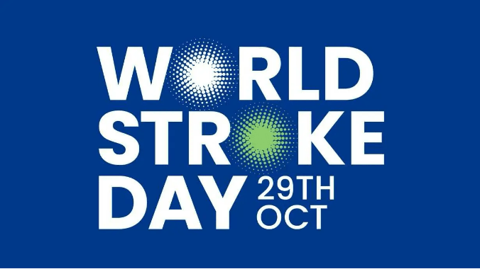
World Stroke Day 29 October

World Stroke Day

When someone has a stroke, every second is crucial. The longer it takes to receive treatment, the more likely it is that damage to the brain will occur.

“The mantra is ‘time is brain’. “The sooner they get treatment, the better patients do.”

World Stroke Day is recognized each year on Oct. 29. The aim is to teach the public about stroke risk factors and stroke prevention, and to raise awareness about the warning signs of stroke so people recognize when a loved one may be having a stroke and can take action.

What is Stroke?

A stroke occurs when a blood vessel bringing blood and oxygen to the brain is interrupted or ruptures (bursts) and brain cells don’t get the flow of blood that they need. Deprived of oxygen, nerve cells can’t function and die within minutes. When nerve cells do not function, the part of the body they control can’t function either. The devastating effects of stroke are often permanent because dead brain cells can’t be replaced.

Types of Stroke

There are two main types of strokes: ischemic and hemorrhagic. An ischemic stroke happens when there is a loss of blood supply to an area of the brain. A hemorrhagic stroke happens when there is bleeding into the brain when a blood vessel ruptures. Eighty-five percent of all strokes are ischemic.

Blood supply to the brain

Blood vessels that carry blood to the brain from the heart are called arteries. The brain needs a constant supply of blood, which carries the oxygen and nutrients it needs to function. Each artery supplies blood to specific areas of the brain. A stroke occurs when one of these arteries to the brain either is blocked or bursts. As a result, part of the brain does not get the blood it needs, so it starts to die.

Possible Effects of Stroke

Motor and sensory function

The human brain is divided into several areas that control movement and sensory function, or how the body moves and feels. When a stroke damages a certain part of the brain that area may no longer work as well as it did before the stroke. This can cause problems with walking, speaking, seeing or feeling.

Functions of right and left hemisphere of the brain

The left side, or hemisphere, of the brain controls how the opposite (right side) of the body moves and feels, and is responsible for how well we can figure out problems with science, understanding what we read and what we hear people say, number skills such as adding and subtracting, and reasoning. The right side of the brain controls the movements and feelings on the left side of the body and is in charge of how artistic we are, including musical and creative talents.

Stages of normal and blocked artery blood flow

  • Normal artery: Blood flows easily through a clear artery.
  • Blockage: An artery can become blocked by plaque (a fatty substance that clogs the artery) or a blood clot, which reduces blood flow to the brain and may cause a stroke.
  • Blockage cleared: The plaque or blood clot breaks up quickly and blood flow is restored to the brain. This may happen during a TIA or mini-stroke, where brain cells recover with no permanent brain damage.

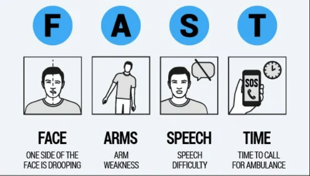
Stroke Symptoms from “Fast to Faster” – देखना, दिखना, हाथ, पैर, बोल, चाल
fast stroke
Fast Stroke Symptoms – देखना, दिखना, हाथ, पैर, बोल, चाल

The acronym FAST (Facial drooping, Arm weakness, Speech difficulties and Time) has been used by the National Stroke Association, American Heart Association and others to educate the public on detecting symptoms of a stroke. FAST was first introduced in the United Kingdom in 1998.

Hindi version of FAST is “Dekhna, dikhna, hath, paer, bol, chal” – could be an easy mnemonic. Any sudden onset disturbance in dekhna, dikhna, hath, paer, bol or chal should raise suspicion of a cerebrovascular event and may indicate prompt medical consultation.
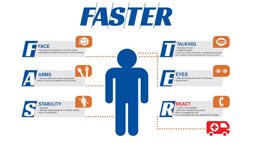
faster stroke symptoms
Faster Stroke Symptoms – देखना, दिखना, हाथ, पैर, बोल, चाल
  • F stands for Face, which refers to drooping or numbness on one side of the face versus the other. Ask the person to smile to make the droop more apparent.
  • A stands for Arms, which refers to one arm being weaker or more numb than the other. Ask the individual to raise both arms up and hold them for a count of ten. If one arm falls or begins to drop, then this could be a sign of a stroke.
  • S stands for Stability, which refers to steadiness on your feet. Sometimes individuals will fall, feel very dizzy or be unable to stand without assistance. Difficulty maintaining balance, trouble walking and loss of coordination are all possible stroke symptoms.
  • T stands for Talking, which refers to changes in speech including slurring, garbled, nonsensical words, or the inability to respond appropriately. Individuals experiencing a stroke may be difficult to understand, or they may have difficulty understanding others. Ask the person to repeat a simple sentence like “The sky is blue.”
  • E stands for Eyes, which refers to visual changes. These visual changes occur suddenly and can include complete vision loss in one eye, double vision, and partial loss of vision in one or both eyes.
  • R stands for React, which is a reminder to call nearby health facility immediately if you recognize any of these symptoms. Call even if the symptoms go away and try to remember when they first began..

Identifying and reacting to stroke symptoms quickly is crucial to achieving proper treatment for an individual experiencing a stroke. Recognizing these “FAST” symptoms and getting treatment quickly can minimize damage to the brain and lessen post-stroke complications.

Diagnosis of Stroke

In addition to a physical examination and laboratory tests, physicians may use a variety of advanced imaging diagnostic tests to diagnose a stroke.

In the emergency room, your doctor or stroke emergency team will:

  • ask you when the symptoms of the stroke started
  • ask you about your medical history
  • conduct a physical and neurological examination
  • order certain laboratory (blood) tests
  • perform imaging tests to help determine what kind of stroke you are having
  • request additional tests that might be needed
  • request additional tests that might be needed, which could include some of the following:

Imaging Tests

CT scan (computed tomography)

An imaging test of the brain that uses radiation to create a picture (like an X-ray) of the brain. It’s usually one of the first tests given to a patient with stroke symptoms as test results give valuable information about the cause of stroke and the location and extent of brain tissue affected.

MRI (magnetic resonance imaging)

An MRI uses a large magnetic field to produce an image of the brain. Like the CT scan, it shows the location and extent of brain injury. The image produced by MRI is sharp and detailed, so it’s often used to diagnose small, deep injuries and may help determine a possible cause of the stroke.

Echocardiogram

An ultrasound imaging procedure used to assess the heart’s function and structures. It can be used to check for conditions such as heart disease, congenital birth defects, heart failure, pericarditis (an inflammation of the lining of the heart) or disease of the valves which might identify the cause of the stroke.

Blood Flow Tests

Carotid artery ultrasound

A carotid artery ultrasound may be ordered if your doctor hears an abnormal sound over your carotid artery caused by disturbances in the blood flow. This diagnostic test takes images of the blood flowing through the arteries and it can detect how severe the narrowing is from plaque buildup.

Cerebral angiography/cerebral arteriography

A cerebral angiography/cerebral arteriography can be performed to diagnose and show the degree of carotid artery stenosis. This test feeds a catheter from your groin, through your aorta and into the carotid artery. An injectable contrast dye is then inserted into the artery while images of the area are captured. This dye allows your doctor to view the arteries in a more enhanced field of view to detect any vessel abnormality. This test is similar to a catheterization to the heart.

Prevention of Stroke

Many strokes can be prevented in the first place by minimizing risk factors. Maintaining a healthy body weight, staying physically active and controlling blood pressure reduce the risk of stroke. Other stroke prevention steps include stopping smoking, eating a healthy diet and managing blood sugar levels.
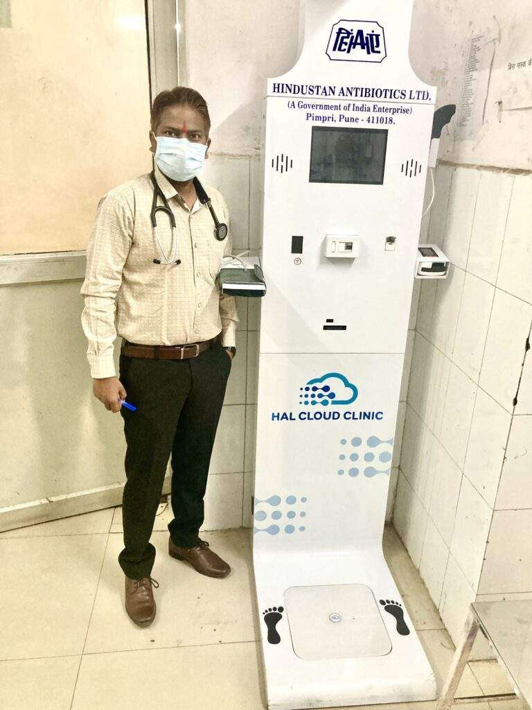
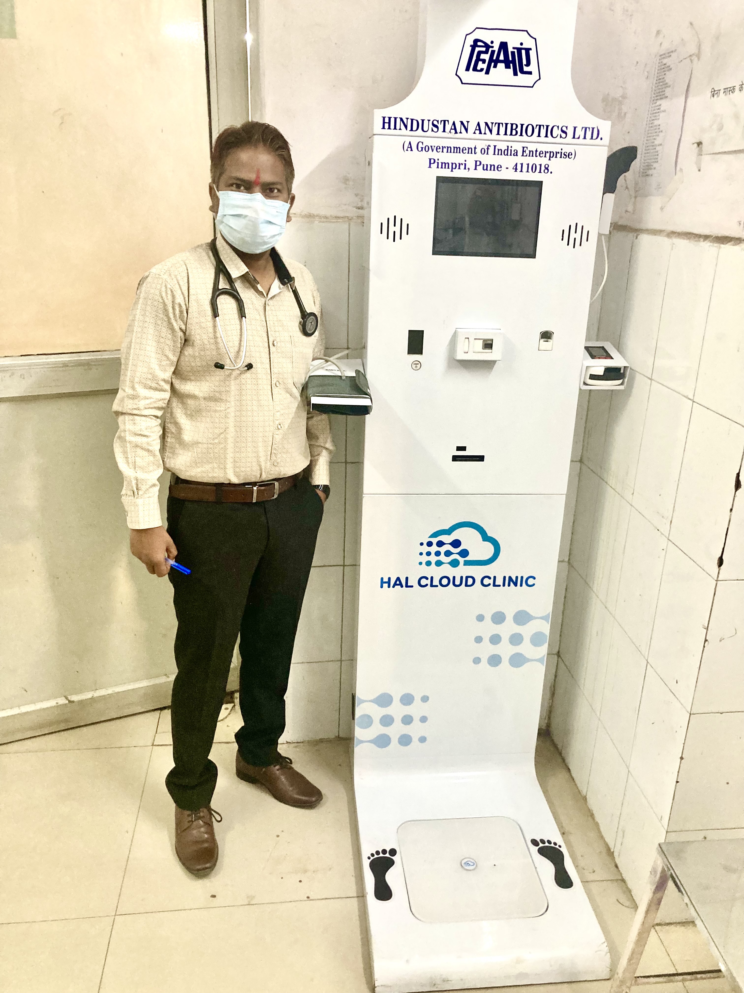
Digital Health ATM Machine from Hindustan Antibiotics Ltd.

No. 1 Digital Health ATM Machine

Digital Health ATM Machine from Hindustan Antibiotics Ltd.

What is Digital Health ATM Machine?

Clinics on cloud health kisosk is an aggregtion of CE/FDA/Medical Grade devices combined with HIPPA complaint backened software which solves problem of basic health awarness checkup.

The company has conducted 100 health machines across the state and for this, it had entered into an agreement with ‘Clinics on Round’, firm. Interestingly, this machine can be handled by anyone competently.

People will be able to get their full body check-up done in 10 minutes at a Health ATM. These health ATMs will be installed in parks, markets, hospitals and such places where there is more movement of people. Tests for dengue, malaria, HIV, typhoid will be done.

Efficiently conducting the tests to check corona infection without any help from a doctor, the Hindustan Antibiotics Company Limited (HAL) has come up with the ‘Health ATM’, a digital machine which was one of its kinds in India. The Pimpri based company HAL through this machine will conduct 22 different tests and will give the results within five minutes stating whether you are fit or unfit.

Inbuild Devices in Digital Health ATM Machine: The advanced technology

  • Blood pressure
  • Glucometer
  • Thermometer
  • Oximeter
  • Hemoglobinometer
  • Digital Height Sensor
  • Body fat analyser etc.

Total 22 parameters measured in digital health ATM machine:

BMI, BMR, Body fat, Body water, Bone mass fat, Free weight, Muscles mass, Protein, Skeletal muscles subcutaneous mass, Visceral fat, Weight, Physic rating, Metabolic age, Health score, Height, BP, Blood sugar level, Pulse, SPO2, Body temperature and Haemoglobin.

What all can be done

  • To install 100 health ATS on an experimental basis
  • 10,000 machines to be produced in the first phase
  • To be given for emergency services
  • Immediate use in the high-risk area
  • To store the health information digitally
  • HIPPA (Health insurance portability and accountability act 1996) 

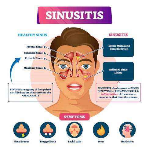
sinusitis

Sinusitis (Rhinosinusitis)

sinusitis
Sinusitis Symptoms
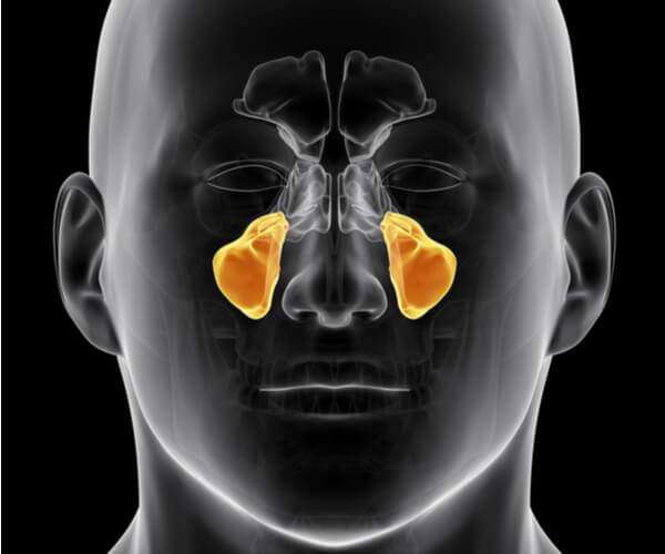
maxillary sinus
Maxillary sinus location

What is Sinusitis?

An infection of the sinuses is known as acute sinusitis. Rhinosinusitis is often a better word since the sinus passageways and nasal passages are connected. Acute rhinosinusitis is a frequent diagnosis, resulting in a significant amount of yearly healthcare costs and plenty of visits to primary care facilities. Additionally, it is a typical justification for prescribing antibiotics. 

It can be brought on by bacterial, viral, or fungal infections, with viral infections being the most frequent. Antibiotics are frequently overprescribed in the treatment of this ailment, so it’s crucial to understand how to correctly examine a sinusitis patient and determine when antibiotics are necessary. In accordance with the recommendations made by several societies, this article covers the causes of rhinosinusitis and when antibiotic treatment in the management of this illness might be appropriate.

Different types of rhinosinusitis may be separated into the following categories based on consensus opinions :

  1. Acute – Signs and symptoms for fewer than four weeks.
  2. Subacute – It takes between four and twelve weeks for symptoms to subside.
  3. Chronic – Enduring symptoms for more than 12 weeks.
  4. Recurrent – Four episodes lasting fewer than four weeks, with full symptom relief between each episode.

Occurrence 

One out of every five antibiotic prescriptions for adults is for acute rhinosinusitis, making it the sixth most prevalent cause for an antibiotic prescription. 6 to 7 per cent of children with respiratory symptoms are affected by acute rhinosinusitis.

Approximately,

  1. 16% of individuals are diagnosed yearly with ABRS. Given the clinical nature of this diagnosis, an overestimation is possible.
  2. An estimated 0.5 to 2.0% of viral rhinosinusitis (VRS) in adults.
  3. And 5 to 10% of children will progress to bacterial infections.

Causes 

  1. The most prevalent cause of acute rhinosinusitis is viruses.
  2. The microorganisms responsible for viral rhinosinusitis (VRS) include rhinovirus, adenovirus, influenza virus, and parainfluenza virus.
  3. Streptococcus pneumonia (38%) is the most prevalent cause of acute bacterial rhinosinusitis (ABRS), followed by Haemophilus influenzae (36%) and Moraxella catarrhalis (16%).
  4. Rarely, fungal infections may also cause acute rhinosinusitis, although this is virtually only seen in immunocompromised people.
  5. It is crucial to distinguish between acute invasive fungal sinusitis (IFS) and allergic fungal sinusitis (AFS), which manifests in immunocompetent people as a mass-like lesion filling a sinus canal and often causes persistent symptoms.

How to assess the patient?

1. Clinical evaluations often identify acute rhinosinusitis. The most sensitive and specific “cardinal” symptoms for acute rhinosinusitis are purulent nasal discharge accompanied by nasal obstruction or face pain/pressure/fullness. This must be determined particularly from people who report “headache” as a general complaint. Facial pressure is a symptom of sinusitis, but the headache is not (with the rare exception of sphenoid sinusitis, which may manifest as an occipital or vertex headache and is often persistent). The observant doctor must gather this information from the patient in order to ascertain the patient’s precise symptoms.

2. ABRS may be diagnosed if cardinal symptoms continue beyond ten days or if they intensify after an initial period of recovery (“double worsening”). Acute rhinosinusitis is accompanied by cough, weariness, hyposmia, anosmia, maxillary dental discomfort, and ear fullness or pressure. Mucopus coming from the osteomeatal complex may be detected by anterior rhinoscopy, or it may be proven by formal endoscopic rhinoscopy in the clinic.

3. The clinical manifestation of ABRS differs somewhat across children. Children are more likely to appear with fevers, in addition to the 10-day length, cardinal symptoms, and “double worsening.” Initial nasal discharge may be watery, then become purulent. Approximately 80% of acute bacterial sinusitis is preceded by an upper respiratory infection.

4. The severity of the symptoms suggests a bacterial origin. At the onset of the disease, these symptoms include high fevers (above 39 C or 102 F) accompanied by purulent nasal discharge or face discomfort for three to four consecutive days. Generally, viral diseases resolve within three to five days.

Antibiotic resistance issues must also be taken into account. These include:

  • Antibiotic usage during the past month
  • Hospitalisation within the preceding five days
  • Healthcare profession
  • Local antibiotic resistance trends are known to local healthcare providers

Finally, it is important to determine whether a patient is at increased risk. Included among these attributes are:

  • Comorbidities (i.e., cardiac, renal, or hepatic disease)
  • Immunocompromised states
  • Age under 2 years or over 65 years

5. In immunocompromised patients, acute fungal rhinosinusitis is often accompanied by fevers, nasal blockage or bleeding, and face discomfort; however, it may also be asymptomatic. Refractory or severe symptoms should urge investigation of this diagnosis in immunocompromised patients.

How to diagnose the condition?

Clinical evaluations often identify acute rhinosinusitis. It is essential for the doctor to differentiate between VRS and ABRS in order to guarantee the appropriate use of antibiotics.

Local resistance patterns and prevalence of penicillin non-susceptible S. pneumoniae warrants clarification.

The conventional diagnostic criteria for adult rhinosinusitis include the presence of at least two significant symptoms or one major symptom plus two or more mild symptoms. In youngsters, the requirements are the same, with the exception of a greater focus on nasal discharge (rather than nasal obstruction).

Principal Symptoms :

  • Purulent anterior nasal discharge
  • Fever (for acute sinusitis only)
  • Purulent or discoloured posterior nasal discharge
  • Facial congestion or fullness
  • Nasal congestion or obstruction
  • Facial pain or pressure
  • Hyposmia or anosmia

Mild features:

  • Headache
  • Ear pain or pressure or fullness
  • Halitosis
  • Dental pain
  • Cough
  • Fever (for subacute or chronic sinusitis)
  • Fatigue

Here is some clinical advice for telling ABRS from VRS:

  1. Duration of symptoms exceeding 10 days.
  2. At the onset of the disease, a high temperature (above 39 C or 102 F) is accompanied by purulent nasal discharge or face discomfort for three to four consecutive days.
  3. Increase in symptom severity within the first 10 days.

In general, routine laboratory testing is unnecessary. Evaluations for cystic fibrosis, ciliary dysfunction, and immunodeficiency should be considered for chronic, recurring, or persistent rhinosinusitis. Some data suggest that a high ESR and CRP may indicate a bacterial infection.

The gold standard is the culture of endoscopic aspirates with more than or equal to 10 CFU/mL. However, this is not required for ABRS diagnosis and is not performed in the great majority of instances. Due to their weak connection with endoscopic aspirates, nasal and nasopharyngeal cultures are of little value. Referral for endoscopic aspiration may be beneficial for individuals with resistant infections or numerous antibiotic sensitivities.

Imaging is rarely indicated for acute sinusitis unless there is clinical concern for a complication or alternate diagnosis. Plain sinus films are often ineffective in identifying inflammation. They may display air-fluid levels. However, this does not aid in distinguishing between viral and bacterial etiologies. If a complication or other diagnosis is suspected, or if the patient has repeated acute infections, sinus CT imaging should be performed to evaluate for bone, soft tissue, dental, or other structural abnormalities, as well as chronic sinusitis.

These should be attained after an acceptable course of therapy. CT scans of the sinuses may reveal air-fluid levels, opacification, and inflammation. Over 5 mm of thicker sinus mucosa is symptomatic of inflammation. Additionally, it can efficiently evaluate bone deterioration or disintegration. However, these data are not useful for distinguishing between viral and bacterial etiologies.

MRI provides more information than sinus CT when evaluating soft tissue or illuminating a malignancy. Consequently, MRI may be useful for determining the severity of problems in situations involving ocular or cerebral extension.

How to manage the condition?

Antibiotic medication or a period of cautious waiting may be used to treat ABRS, provided that reliable follow-up is assured. There are minor differences in the guidelines of various expert committees.

The amended 2015 American Academy of Otolaryngology Adult Sinusitis guideline suggests amoxicillin with or without clavulanate for 5 to 10 days as first-line treatment for the majority of people. Failure of treatment is determined if symptoms do not improve or worsen within seven days.

The Infectious Disease Society of America Guidelines for Acute Bacterial Rhinosinusitis prescribe amoxicillin with clavulanate for 10 to 14 days in children and 5 to 7 days in adults as first-line treatment. Failure of treatment is determined if symptoms do not improve within 3 to 5 days or worsen within 48 to 72 hours.

the American Academy of Pediatrics Clinic Practice Guideline for the diagnosis and management of acute bacterial sinusitis in children aged 18 years recommended amoxicillin with or without clavulanate as first-line treatment. Uncertainty surrounds the length of therapy, although their recommendation was to continue treatment for a further seven days after symptoms disappear.

If, after 72 hours of therapy, symptoms do not improve or worsen, the treatment has failed. If the patient cannot accept oral fluids, ceftriaxone 50 mg/kg may be administered. If the patient can tolerate oral fluids the next day and improves, he or she may then begin an oral antibiotic regimen. To effectively address beta-lactamase-producing bacteria, a separate article recommends amoxicillin with clavulanate as the first treatment for children.

Adding clavulanate or prescribing high-dose amoxicillin (90mg/kg/day vs 45mg/kg/day) in children is determined by local antibiotic resistance trends, the patient’s risk level, risk factors for antibiotic resistance, and the severity of symptoms.

A third-generation cephalosporin with clindamycin (for enough coverage of non-susceptible S. pneumoniae) or doxycycline might be therapeutic options for penicillin-allergic individuals. The effectiveness of third-generation cephalosporins alone against S. pneumoniae is inconsistent. Fluoroquinolones might also be explored, although they have a greater incidence of adverse effects. In youngsters, doxycycline and fluoroquinolones should be administered with more care. S. pneumoniae and Hemophilus influenzae have elevated levels of resistance to second-generation cephalosporins, trimethoprim/sulfamethoxazole, and macrolides.

There is also evidence that antibiotic medication does not always reduce the duration of symptoms or the risk of complications in adults. Many instances of ABRS may also resolve spontaneously within two weeks.

Symptomatic therapies 

Clinicians may give symptomatic therapies, but generally, there is a lack of conclusive data. In guidelines, nasal steroids and nasal saline irrigation are the most popular suggestions. By lowering mucosal oedema, intranasal steroids may assist in relieving the blockage. A limited number of clinical studies suggested that greater dosages of intranasal corticosteroids may reduce the period of symptom remission by two to three weeks. Additionally, nasal saline irrigation may aid in reducing blockage. Due to their ability to thicken nasal secretions, antihistamines are not recommended unless there is a definite allergic component.

Be aware of potential complications

Complications are uncommon, occurring in around one out of every thousand cases. Infections of the sinuses may extend to the orbit, bone, and cerebral cavities. 80% of orbitocranial problems manifest in the orbit. These problems may be associated with substantial morbidity and death. Due to the very thin ethmoid bone that divides infections from the ethmoid from the orbit, the orbit is the most likely location.

Prognosis

Most cases of acute bacterial rhinosinusitis are viral. The vast majority of cases are either self-limiting or efficiently treatable with antibiotics. In immunocompromised individuals, invasive fungal rhinosinusitis is an uncommon but severe type of illness. It is related to a high risk of morbidity and death.

Shingles (Herpes Zoster)

Painful Shingles (Herpes Zoster) – 2 Vaccines

Shingles (Herpes Zoster)
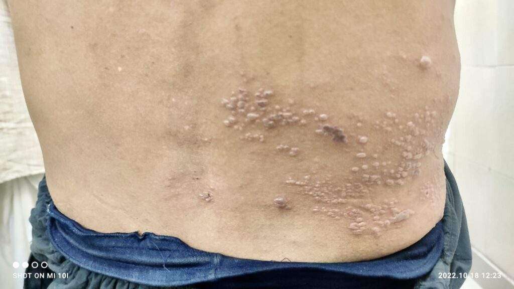
a patient with shingles (herpes zoster infection)

What is shingles?

It is a viral infection that causes an outbreak of a painful rash or blisters on the skin. It’s caused by the varicella-zoster virus, which is the same virus that causes chickenpox. The rash most often appears as a band of rashes or blisters in one area of your body.

Where does it come from?

When you have chickenpox as a child, your body fights off the varicella-zoster virus and the physical signs of chickenpox fade away, but the virus always remains in your body. In adulthood, sometimes the virus becomes active again. This time, the varicella-zoster virus makes its second appearance in the form of shingles.

How common is shingles?

About 1 million cases are diagnosed every year. The risk of shingles increases as you get older, with about half the cases occurring in people over the age of 50. Shingles develops in about 10% of people who have had chickenpox at an earlier time in their lives.

Who is at risk for getting shingles?

People who have had chickenpox who are more likely to develop shingles include those:

  • With a weakened immune system (such as people with cancer, HIV, organ transplant recipients or those receiving chemotherapy).
  • Over the age of 50.
  • Who have been ill.
  • Who have experienced trauma.
  • Who are under stress.

The chickenpox virus doesn’t leave your body after you have chickenpox. Instead, the virus stays in a portion of your spinal nerve root called the dorsal root ganglion. For the majority of people, the virus stays there quietly and doesn’t cause problems. Researchers aren’t always sure why the virus gets reactivated, but this typically occurs at times of stress.

Can you get shingles more than once?

Yes, you can get shingles more than one time. One of the biggest myths about shingles is that it can only happen once. This isn’t true. You can have more than one episode. If you get shingles again, you usually don’t get the rash in the same place.

What are the symptoms?

Early symptoms may include:

  • Fever.
  • Chills.
  • Headache.
  • Feeling tired.
  • Sensitivity to light.
  • Stomach upset.

Other signs and symptoms that appear a few days after the early symptoms include:

  • An itching, tingling or burning feeling in an area of your skin.
  • Redness on your skin in the affected area.
  • Raised rash in a small area of your skin.
  • Fluid-filled blisters that break open then scab over.
  • Mild to severe pain in the area of skin affected.

How long does a shingles outbreak last?

It can take three to five weeks from the time you begin to feel symptoms until the rash totally disappears.

  1. First, a few days before the rash appears, you may feel pain in an area on your skin. The pain is described as itching, burning, stabbing or shooting. This usually happens before the rash comes.
  2. Next, the raised rash appears as a band or a patch, usually on one side of your body. The rash usually appears around your waistline or on one side of your face, neck, or on the trunk (chest/abdomen/back), but not always. It can occur in other areas including your arms and legs.
  3. Within three to four days, the rash develops into red, fluid-filled, painful, open blisters.
  4. Usually, these blisters begin to dry out and crust over within about 10 days.
  5. The scabs clear up about two to three weeks later.

Do you always get the typical rash if you have shingles?

Occasionally, some people don’t get a rash. If you have any of the other symptoms (even without a rash), see your healthcare provider sooner rather than later. There are effective treatments you can take early for shingles. Even if you don’t have shingles, seeing your healthcare provider will help you get your condition diagnosed and treated.

Why does shingles appear mostly on one side or in one area of your body?

The virus travels in specific nerves, so you will often see shingles occur in a band on one side of your body. This band corresponds to the area where the nerve transmits signals. These rash stays somewhat localized to an area. It doesn’t spread over your whole body. Your torso is a common area, as is your face.

Is shingles contagious?

Someone can’t spread shingles to another person, but they can spread chickenpox. The varicella-zoster virus is spread through direct skin-to-skin contact with the fluid that oozes from the blisters. Shingles is rarely spread by breathing in the varicella-zoster virus the way airborne viruses are spread. If your rash is in the blister phase, stay away from those who haven’t had chickenpox or the chickenpox vaccine and keep your rash covered.

How long are you contagious if you have shingles?

If you have such lesion, you’re contagious until the rash is dried and crusted over. The varicella-zoster virus can only cause chickenpox in someone who has never had chickenpox or hasn’t been vaccinated against chickenpox.

How is shingles diagnosed?

It can be diagnosed by the way the rash is distributed on your body. The blisters of a shingles rash usually appear in a band on one side of your body. It may also be diagnosed in a laboratory using scrapings or a swab of the fluid from the blisters.

How is shingles treated?

There is no cure for shingles but there are treatments for managing the symptoms.

Antiviral medications

These drugs may ease the discomfort and make the symptoms stop sooner, particularly if you start them within 72 hours of the first sign of shingles. They may also help prevent the pain that can happen months and years later, called post-herpetic neuralgia. These medications include:

  • Acyclovir.
  • Famciclovir.
  • Valacyclovir.

Over-the-counter pain medications

These medications include the following and may be effective in relieving pain:

  • Acetaminophen.
  • Ibuprofen.

Other medications

Antibacterial drugs may be prescribed if you develop a bacterial infection due to the shingles rash. Anti-inflammatory drugs like prednisone may be prescribed if shingles affects your eyes or other parts of your face.

If you have more than one area of blisters, what can you expect if you go to the hospital?

It’s important to note that most people with shingles don’t need to be in a hospital, but if you do:

  • You’ll be in a contact isolation room.
  • The door will be kept closed.
  • A sign on your door will remind people who have never had chickenpox or the vaccine not to enter.
  • The sign will also remind staff to wear gowns and gloves when entering the room.

If you have shingles in only one area of your body that can’t be kept covered, what can you expect for your hospital stay?

  • You will be in a contact isolation room.
  • The sign on the door will remind staff to wear gowns and gloves when entering the room.

What are the complications of shingles?

After the shingles rash has disappeared, you might continue to have nerve pain in that same area. Post-herpetic neuralgia can last for months or years and become quite severe.

More than 10% of people who get shingles develop post-herpetic neuralgia. Researchers don’t know why some people get post-herpetic neuralgia and others don’t. It may be that nerves become more sensitive or that the virus may be invading and damaging the central nervous system.

Other complications include:

  • Other types of nerve issues like numbness or itching.
  • A bacterial infection of the shingles rash.
  • Eye and ear inflammation if the rash is near these organs.

Is shingles dangerous or even fatal?

If shingles involves your eye, it can lead to blindness. In rare cases, shingles can lead to hearing problems, pneumonia, inflammation of the brain (encephalitis) and even death.

How is post-herpetic neuralgia treated?

Treatments include lotions or creams (such as lidocaine or capsaicin) and/or other medications not specifically used for pain, such as antidepressants or drugs for epilepsy. Regular pain relievers are not usually effective for this type of pain.

If your pain doesn’t lessen, you might try therapies like nerve blocks or steroid injections near the area where the nerves exit the spine. Your provider might suggest an implantable nerve stimulator device for severe, ongoing pain that hasn’t responded to other treatments.

Is a vaccine available to prevent shingles?

Two vaccines are available in the market to reduce your chance of developing shingles and post-herpetic neuralgia. One vaccine, Zostavax®, has been available since 2006. The second vaccine, Shingrix®, has been available since 2017. Shingrix is recommended as the preferred vaccine by the Advisory Committee on Immunization Practices, a group of medical and public health experts.

Shingrix (recombinant zoster vaccine) is given as a two-dose shot in your upper arm. You should receive the second dose (shot) two to six months after receiving the first. Shingrix has been shown to be more than 90% effective in preventing shingles and postherpetic neuralgia. Its effectiveness remains above 85% for at least four years after receiving the vaccine.

Who should be vaccinated with Shingrix?

The Shingrix vaccine is recommended for those 50 years of age and older who are in good health.

You should get the Shingrix vaccine even if:

  • You’ve had shingles already.
  • You’ve been previously vaccinated with Zostavax (a live zoster vaccine). If you’ve been vaccinated with Zostavax, wait at least eight weeks before getting vaccinated with Shingrix.
  • You don’t know for sure if you’ve ever had chickenpox.

Ask your healthcare provider, who knows your entire health history if getting this vaccine is right for you.

Who should not be vaccinated with Shingrix?

You shouldn’t receive the Shingrix vaccine if you:

  • Have ever had a severe allergy to this vaccine or any ingredient in this vaccine.
  • Are breastfeeding or pregnant.
  • Currently have shingles.
  • Are ill and have a high fever.
  • Have tested negative for immunity to varicella-zoster virus (get the chickenpox vaccine instead).

Ask your healthcare provider if the benefits of getting the vaccine outweigh any potential risks.

What serious side effects should you watch for after getting the Shingrix vaccine?

Serious side effects from vaccines are extremely rare. However, call or go to the nearest emergency room right away if you experience any of the following within minutes to hours after receiving Shingrix:

  • Swelling of your face or throat.
  • Difficulty breathing.
  • Hives.
  • Fast heartbeat.
  • Dizziness, lightheadedness, weakness.

If you’ve had shingles recently, how long should you wait before getting the Shingrix vaccine?

You can get the Shingrix vaccine any time after the shingles rash has gone away.

When is it safe to return to work if you have shingles?

You can return to work when you feel well enough to return and you’re no longer contagious. This means that your blistered rash has dried up and scabbed over. This usually takes up to 10 days from the time the rash first appears.

Are there natural ways to boost your immune system to help lessen the chances of developing shingles?

Stress is a risk factor for developing it, so limiting your stress can be helpful. Try meditation, yoga or other relaxation methods.

Other things you can do include:

  • Eat a healthy diet.
  • Maintain a healthy weight.
  • Exercise regularly.
  • Aim for seven to nine hours of sleep each night.
  • Don’t smoke or use tobacco products.

These are all tips for an overall healthy lifestyle, not just for reducing your chance of getting shingles.

What is the difference between herpes zoster and varicella-zoster?

Herpes zoster is simply another medical name for shingles. Varicella-zoster is the virus that causes both shingles and chickenpox.

A note from Bhavishya Clinic+

If you’ve had chickenpox, you’re at risk of developing shingles later in life. It causes a rash that is contagious and painful. The disease can have serious complications. The best thing you can do to reduce your risk is to get the shingles vaccine. The vaccines are safe and effective.

Three-main-types-of-urinary-catheters

3 Main Types Of Urinary Catheters

Three-main-types-of-urinary-catheters
Urinary catheter

What is a Urinary Catheter?

A urinary catheter is a medical device made from a thin, hollow tube that can be inserted through the urethra or through a small opening in the abdomen, in order to drain urine from the bladder. Male external catheters, though they share a similar name, are shaped like a condom and are designed to slip over the penis rather than being inserted into the body.

How does a urinary catheter work?

The hollow tube has openings at the end, so that when it is inserted into the bladder, the urine will naturally flow out through the tube into a collection bag or the toilet. Male external catheters have an adhesive that creates a leak-proof seal around the penis with an opening at the end so the urine will flow out the front, through a tube and into a collection bag.

How is the urinary catheter inserted?

Indwelling catheters will be inserted by a medical professional through the urethra or though a small opening in the abdomen in the case of suprapubic catheters. Intermittent catheters are inserted into the urethra and this can be done by yourself at home, or by a nurse or caregiver.

How do urinary catheters stay in?

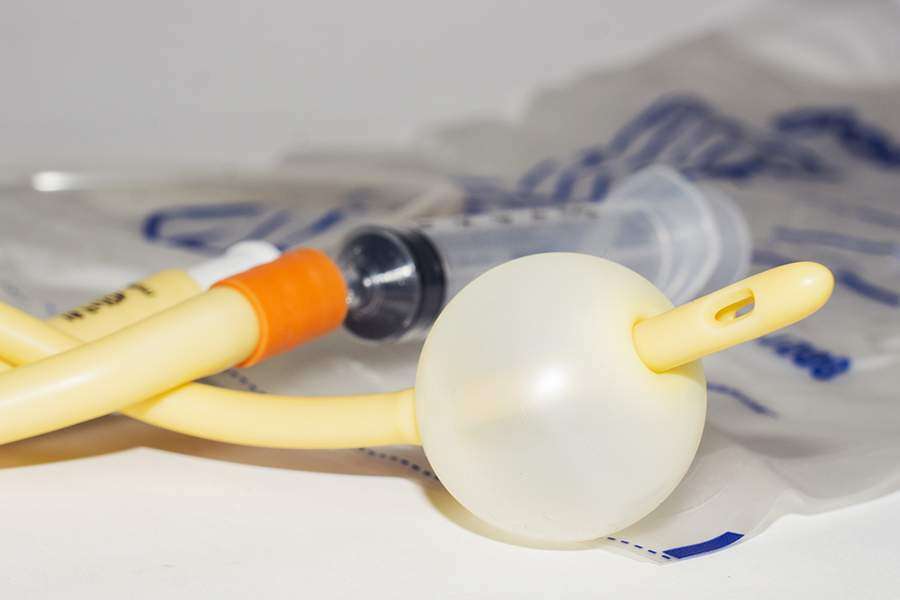
Once an indwelling catheter is inserted into the bladder, an area near the end of the catheter can be inflated with sterile water, like a balloon. This small balloon is much bigger than the urethra so it holds the catheter in place, preventing it from slipping out and still allowing urine to drain through the tube in the middle of the balloon. The balloon can be deflated when the catheter is removed. Keep in mind, not all catheters stay in. Intermittent catheters are inserted only when the bladder is full, quickly draining the bladder and then removed when the bladder has emptied.

Why are urinary catheters used?

There are a great variety of diagnoses that would result in the need of a catheter. Sometimes a catheter is needed only temporarily during hospitalization. Often times they are used permanently when the bladder does not function normally or when the bladder cannot be controlled. Catheters may be used as a solution for bladder retention (inability to empty the bladder) or bladder incontinence (inability to control the bladder).

What are the different types of urinary catheters?

There are three main types of catheters: indwelling catheters, intermittent catheters, and external catheters for men. Catheters come in different materials, with differently shaped tips based on the user’s needs. More information on the types of catheters can be found below.

Indwelling urinary catheters

Indwelling catheters are designed to be inserted through the urethra (Foley catheter) or a small opening in the abdomen (suprapubic catheter). Indwelling catheters are designed to stay inside the body to continuously drain the bladder. Indwelling catheters are inserted by a medical professional and can be used long term, with the catheter being changed monthly.

Intermittent urinary catheters

Intermittent catheters are also known as “in and out” catheters because they are inserted and removed around 5 times a day. When inserted, they are designed to drain the bladder all at once and then be removed. The insertion of an intermittent catheter can be done in the home once the user learns the technique and has had a little practice. There are different variations of intermittent catheters, explained below.

  • Straight intermittent catheter: This catheter can be made from latex or silicone, is generally used with lubricant, and has a funnel at the end to drain into the toilet. This is the standard intermittent catheter.
  • Closed system catheters: These come pre-lubricated and are attached to a collection bag. The system is sterile and touch-free, which can help prevent infections. The urine drains into the collection bag and then is disposed of, allowing for convenient catheterization when there is not a restroom nearby.
  • Hydrophilic coated catheters: Catheters coated with a slippery surface that is activated with water. This eliminates the need for a separate lubricant and may reduce irritation to the urethra for frequent catheter users.

Male External Catheters, a.k.a. Condom Catheters

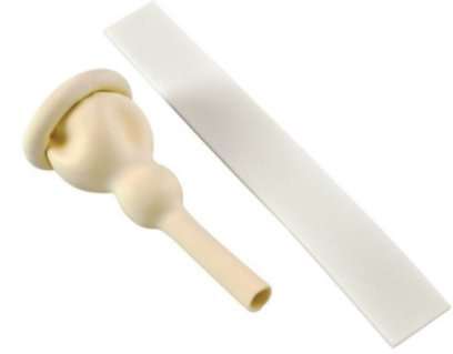
Male external catheters are also known as condom catheters, Texas catheters, or “gizmos.” These are different from the other catheters because they are external, so they are not inserted into the body. These catheters fit like a condom, so they can be slipped over the penis in the same way. Usually they will have an adhesive that seals against the skin of the penis, preventing leakage.

A tube connects to the front of the catheter, allowing the urine to flow out into a collection bag. Male external catheters are generally used for continuous draining of the bladder. They can be used at home with proper training, or a medical professional can change it for you. Male external catheters should be changed daily.

condom catheter
condom catheter
Breathe easy this diwali

BREATHE EASY THIS DIWALI 2022

Breathe easy this diwali

Measures to breathe easy this Diwali?

Breathe easy this Diwali which is arguably India’s biggest festival which is synonymous with lights, lamps, colors and of course, firecrackers. However, this is also the time when air quality deteriorates drastically. The air is chocked with various forms of pollutants with the problem compounded by pollution caused by the increased movement of vehicles.

Patients visiting to the hospitals with complaints of severe respiratory disorders during Diwali festival have been increasing for the last five years. These include asthma, ARDS, and exacerbations of chronic lung disorders (COPD), all associated with breathing in polluted and toxic air. The problem of pollution is because of the effects of fireworks in Diwali. It is more severe in cities as the growing number of vehicles adds to the problem.

Air quality takes a deep plunge as the problem gets compounded. Depending on the size of these particles, they cause a variety of illnesses ranging from mild temporary irritation to serious chronic diseases such as cancer.

Particles with a diameter of 10 micrometers (PM 10) and less are considered the most harmful as they are small enough to evade the body’s filters and pass through the entire respiratory system. Travelling from the upper respiratory tract which consists of the nose, pharynx and larynx, the smaller particles end up accumulating in the lower respiratory tract (bronchioles, alveolar ducts, and alveolar sacs) where gas exchanges occur. These are eventually removed by the macrophages of the immune system.

When there is an increased number of particulate matter in the air along with gases, the chances of COPD exacerbations increase. Certain groups of people are particularly susceptible, such as:

Children – Children spend a lot of time playing outdoors and this is the reason why they run a high risk of getting affected by smog. Regular exposure to air pollution (smog) can cause asthma and even other harmful respiratory ailments.

People who do outdoor activities – Anyone who works outdoor is more susceptible to air pollution. Regular exposure and can deteriorate their health causing several respiratory diseases.

People with Asthma & Respiratory disease – People with asthma or chronic respiratory diseases are at a high risk of health hazards caused by smog. They will experience the adverse effects of air pollution much sooner compared to others. Continuous exposure to smog can aggravate lung diseases, heart disease and stroke.

Elderly People – Senior citizens have a weak immune system. Owing to this condition, elderly people are at increased risk of getting affected by air pollution, which can cause cardiac arrhythmias and heart attacks, asthma attacks and many more.

Dry hacking coughs, shortness of breath, chest tightness, wheezing, nasal complaint along with sneezing are some of the common complaints of patients during this time. Air pollution not only affects your lungs but other organs. Thus, it is important to know the health hazards during Diwali time and take precautions accordingly.

  1. Upper Respiratory Tract Infection: The Post-Diwali period usually witnesses a surge in the number of people with complaints of irritation in the eyes and throat, dry cough and fever. While last year’s smog lingered in the atmosphere, many people who otherwise remain healthy, also experienced irritation in eyes and chest and a general feeling of suffocation.
  2. Bronchitis: The deadly chemicals used in crackers release fumes and gases that can lead to the inflammation of the bronchial tubes leading to acute attacks.
  3. Asthma: The condition of asthma patients usually exacerbates during this period because of the toxic fumes in the air.
  4. COPD: The risk of Chronic Obstructive Pulmonary Disease (COPD) increases during festivals like Diwali when the level of suspended particulate matter becomes high in the air. It is characterized by prolonged cough with excess sputum.

Persons suffering from asthma, bronchitis and other respiratory ailments should start taking precautions several days before the festive season. Also, it is advisable for affected populations to avoid outdoor physical activities and wait until the levels of fine particles decrease. People suffering from Asthma, if it cannot be avoided should use a N95 face mask, which filters out at least 95% of airborne particles.

A normal surgical mask is of no use as pollutants can pass through it. It is important to keep inhalers handy. If one experiences breathlessness then it is advisable to move away from smoggy surroundings and sit in a room with the doors and windows shut and the air-conditioning on. Patients are supposed to continue their treatment for chronic lung disease regularly and consult pulmonologist at regular intervals. It is important to keep an inhaler or nebulizer at hand to dilate your airways. Rush to a hospital if you don’t feel better after inhalation.

Do’s & Don’ts to Stay Safe and breathe easy this Diwali

DOs

  • Use Multi Layered Masks: While going out, ensure that your nose and mouth are covered with a good quality mask to ban the entry of harmful particles in your respiratory tract. If you don’t have a mask, simply use a handkerchief or cloth to cover up.
  • Use Air Purifiers: Air inside closed spaces is more polluted than outside air. It’s advisable to install air purifiers in at homes and offices that allow only a little movement of air.
  • Use Air Purifying Plants: Your home and office must have plenty of plants that purify the air. A noteworthy point here is that there must be a variety of such plants and they must be spaced appropriately.
  • Use Air Quality Checking Apps: Before you go out, check the level of pollution through various Apps. If the air quality is severe, avoid going out at all.
  • Moisturize Your Nostrils: Moisturizing your nostrils helps in restricting the entry of fine particulate matter through your nose.

DON’Ts

  • Morning and Evening Walks/Runs: Air quality is worst in the mornings. Simply avoid morning and evening walks or running outside altogether till air quality improves.
  • Heavy Exercising: Do only light exercises, as heavy exercises require heavy breathing.
  • Eating Fried/Junk Food: Ensure you eat light, healthy, fiber rich and fat free food to feel healthier and fresh.
  • Travelling on two wheelers: Say No to travelling on two wheelers- this can be hazardous for your health.

Human life is above all and good health is a prerequisite of a happy life. We urge everyone to celebrate Diwali responsibly this year- in a way which makes every face glow with a smile and does not put anyone at any kind of health risk.

“Wishing You All, Happy & Safe Diwali”

calorie calculator

Calculate Your Optimal Calories in 2022

Mifflin – St. Jeor Calorie Calculator

Calculate Your Optimal Calories
Imperial
Metric
Basic Information
Activity Level
Your results:
Target calorie intake per day:
0

How many calories do I need?

An acceptable macronutrient distribution range for carbohydrates (45-65% of energy), protein (10-35% of energy), and fat (20-35% of energy; limit saturated and trans fats). These recommendations are broad and meant to cover the needs of many different people with different dietary situations. This is a much-discussed and debated issue, and the current understanding is that there is no one-size-fits-all recommendation. How many calories you consume and what percent of your calories come from each macronutrient can be manipulated to meet your individual needs and goals. However, consuming adequate protein at or above the recommended amount is necessary to maintain lean body mass.

About

The Mifflin-St Jeor equation is a widely used tool to determine the resting metabolic rate [RMR], which is defined as the number of calories burned while the body is in complete rest. RMR is also known as resting energy expenditure [REE]. The equation was developed by MD Mifflin and ST St Jeor and first introduced in a paper published in 1990. We are using this calorie calculator at our Bhavishya Clinic+

There are several equations for measuring RMR, including the most popular Harris-Benedict equation which was developed in 1919 and revised for accuracy in 1984. A comparative study of four predictive equations found that the Mifflin-St Jeor equation is more likely than the other equations to predict RMR to within 10% of that measured.

Formula

Females: (10*weight [kg]) + (6.25*height [cm]) – (5*age [years]) – 161
Males: (10*weight [kg]) + (6.25*height [cm]) – (5*age [years]) + 5

Multiply by scale factor for activity level:
Sedentary *1.2
Lightly active *1.375
Moderately active *1.55
Active *1.725
Very active *1.9

Basal metabolic rate is the amount of energy needed for your body’s physiological functions at rest, such as your breathing, your heart beating, and your brain activity.

This amount of energy required is measured in kilocalories, or often just denoted as calories. Therefore, BMR is your body’s calorie needs at rest with no extra activity.

In order to measure your exact BMR, you need to be in a lab setting and use indirect calorimetry. This measurement is taken under tightly controlled conditions and not easily accessible to most people.

Caloric expenditure measured by indirect calorimetry can then be added to physical activity energy expenditure to calculate total daily energy expenditure.

What is resting metabolic rate (RMR)?

Resting metabolic rate is the total number of calories burned when your body is completely at rest. RMR supports breathing, circulating blood, organ functions, and basic neurological functions. It is proportional to lean body mass and decreases approximately 0.01 kcal/min for each 1% increase in body fatness.

Hunger Vs Appetite

Lastly, take the time to understand some basic differences between hunger and appetite which are outlined below:

Hunger

It is considered a biological response to replenish the body’s energy reserves.

  • Protects us from starvation.
  • Usually triggered by an event occurring below the neckline:
    • Low blood sugar.
    • Empty (growling) stomach.
    • Hormone fluctuations.
    • Need to warm body (hypothermia).
  • Gradual onset, appearing after several hours without food and typically diminishes after eating.
  • It is generally satisfied by almost any food that provides energy (calories).

Appetite

It is considered a desire or interest to eat a specific food.

  • Usually triggered by an event occurring above the neckline consciously or subconsciously:
    • Thoughts, emotions and moods.
    • Social (e.g., happy hour)
    • Cultural (e.g., family)
    • Environmental (e.g., walking into a bakery).
  • More rapid onset and often independent of hunger.
  • Not time-dependent and may persist after eating.
  • Usually only satisfied by a specific food (e.g., sweet, salty) which may then evoke emotions and thoughts afterwards (e.g., pleasure, guilt, shame).

While RMR is an important component of TDEE, an accurate measurement remains elusive for many. Subsequently, we resort to mathematical formulas, but considering their potential errors, the values determined should always be considered a general estimate rather than an accurate value. Given this, there may also be value in including other methods as a guide to avoiding starvation. 

Lastly, while we need to acknowledge the fact that RMR is not entirely controllable, there are some influencing factors we can manipulate and should leverage every opportunity to exploit them.

Cataract

Cataract

Cataract
Cataract
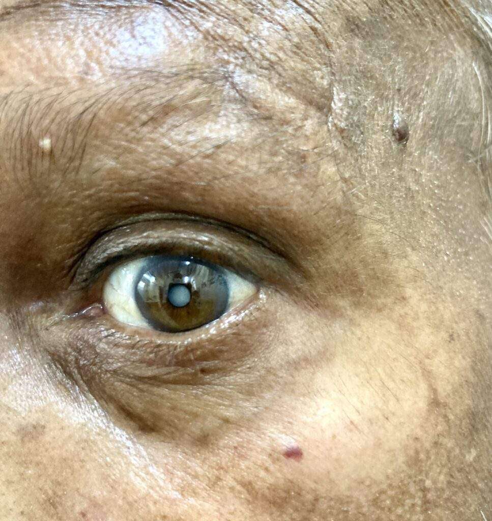
Matyre Cataract
Mature Cataract

Overview

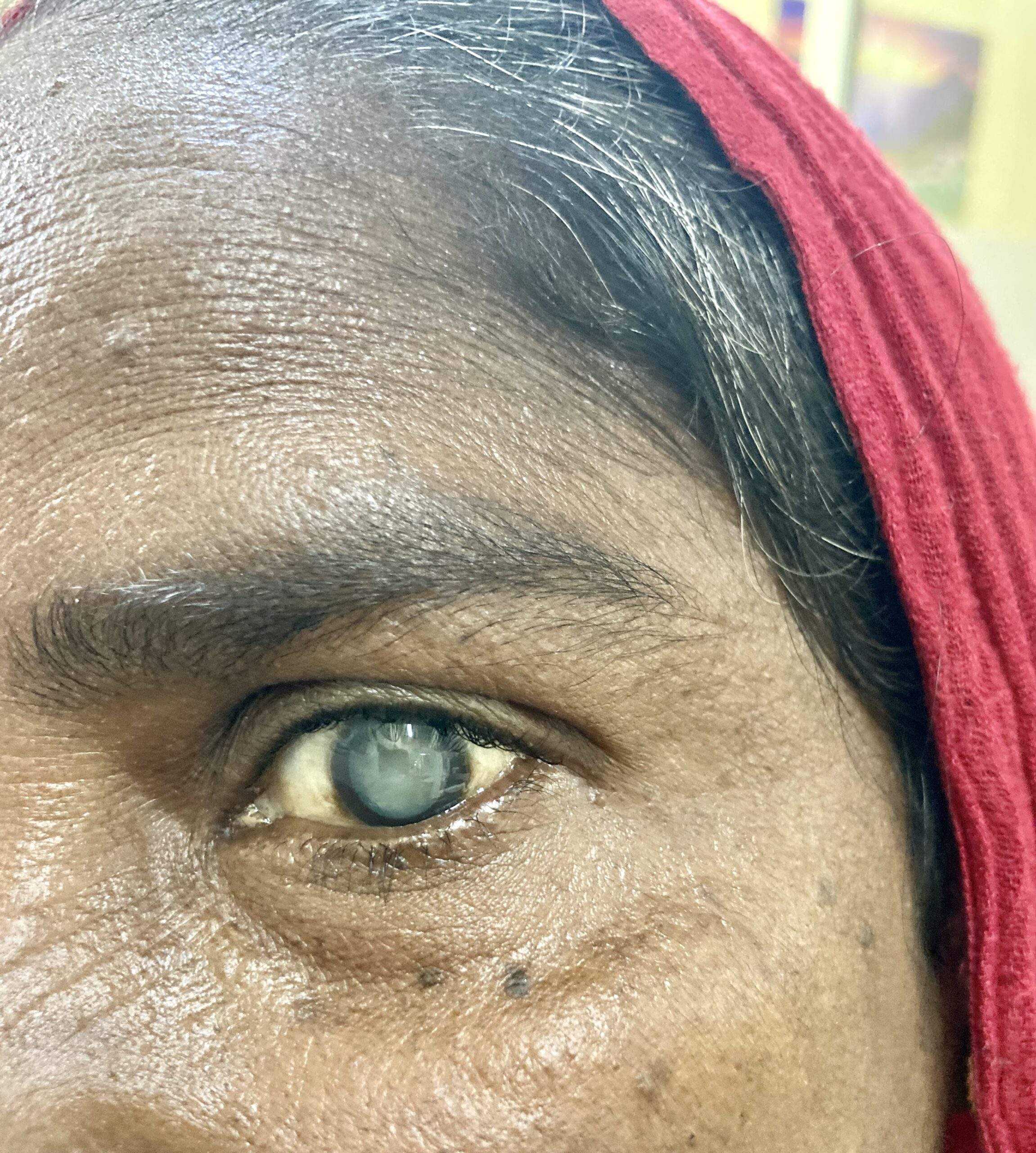
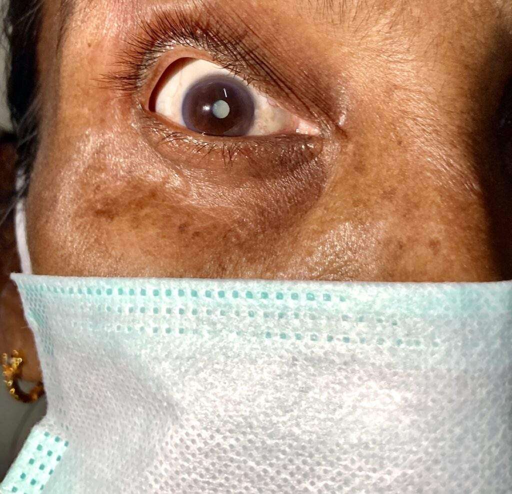
A cataract is a clouding of the normally clear lens of the eye. For people who have cataracts, seeing through cloudy lenses is a bit like looking through a frosty or fogged-up window. Clouded vision caused by cataracts can make it more difficult to read, drive a car (especially at night) or see the expression on a friend’s face.

Most cataracts develop slowly and don’t disturb your eyesight early on. But with time, cataracts will eventually interfere with your vision.

At first, stronger lighting and eyeglasses can help you deal with cataracts. But if impaired vision interferes with your usual activities, you might need cataract surgery. Fortunately, cataract surgery is generally a safe, effective procedure.

Symptoms

Signs and symptoms of cataracts include:

  • Clouded, blurred or dim vision
  • Increasing difficulty with vision at night
  • Sensitivity to light and glare
  • Need for brighter light for reading and other activities
  • Seeing “halos” around lights
  • Frequent changes in eyeglass or contact lens prescription
  • Fading or yellowing of colors
  • Double vision in a single eye
Cataract
Cataract in 62 year old OPD Patient

At first, the cloudiness in your vision caused by a cataract may affect only a small part of the eye’s lens and you may be unaware of any vision loss. As the cataract grows larger, it clouds more of your lens and distorts the light passing through the lens. This may lead to more-noticeable symptoms.

When to see a doctor

Make an appointment for an eye exam if you notice any changes in your vision. If you develop sudden vision changes, such as double vision or flashes of light, sudden eye pain, or sudden headache, see your doctor right away.

Causes

Most cataracts develop when aging or injury changes the tissue that makes up the eye’s lens. Proteins and fibers in the lens begin to break down, causing vision to become hazy or cloudy.

Some inherited genetic disorders that cause other health problems can increase your risk of cataracts. Cataracts can also be caused by other eye conditions, past eye surgery or medical conditions such as diabetes. Long-term use of steroid medications, too, can cause cataracts to develop.

How a cataract forms

A cataract is a cloudy lens. The lens is positioned behind the colored part of your eye (iris). The lens focuses light that passes into your eye, producing clear, sharp images on the retina — the light-sensitive membrane in the eye that functions like the film in a camera.

As you age, the lenses in your eyes become less flexible, less transparent and thicker. Age-related and other medical conditions cause proteins and fibers within the lenses to break down and clump together, clouding the lenses.

As the cataract continues to develop, the clouding becomes denser. A cataract scatters and blocks the light as it passes through the lens, preventing a sharply defined image from reaching your retina. As a result, your vision becomes blurred.

Cataracts generally develop in both eyes, but not always at the same rate. The cataract in one eye may be more advanced than the other, causing a difference in vision between eyes.

Types of cataracts

Cataract types include:

  • Cataracts affecting the center of the lens (nuclear cataracts). A nuclear cataract may at first cause more nearsightedness or even a temporary improvement in your reading vision. But with time, the lens gradually turns more densely yellow and further clouds your vision.As the cataract slowly progresses, the lens may even turn brown. Advanced yellowing or browning of the lens can lead to difficulty distinguishing between shades of color.
  • Cataracts that affect the edges of the lens (cortical cataracts). A cortical cataract begins as whitish, wedge-shaped opacities or streaks on the outer edge of the lens cortex. As it slowly progresses, the streaks extend to the center and interfere with light passing through the center of the lens.
  • Cataracts that affect the back of the lens (posterior subcapsular cataracts). A posterior subcapsular cataract starts as a small, opaque area that usually forms near the back of the lens, right in the path of light. A posterior subcapsular cataract often interferes with your reading vision, reduces your vision in bright light, and causes glare or halos around lights at night. These types of cataracts tend to progress faster than other types do.
  • Cataracts you’re born with (congenital cataracts). Some people are born with cataracts or develop them during childhood. These cataracts may be genetic, or associated with an intrauterine infection or trauma.These cataracts may also be due to certain conditions, such as myotonic dystrophy, galactosemia, neurofibromatosis type 2 or rubella. Congenital cataracts don’t always affect vision, but if they do, they’re usually removed soon after detection.

Risk factors

Factors that increase your risk of cataracts include:

  • Increasing age
  • Diabetes
  • Excessive exposure to sunlight
  • Smoking
  • Obesity
  • High blood pressure
  • Previous eye injury or inflammation
  • Previous eye surgery
  • Prolonged use of corticosteroid medications
  • Drinking excessive amounts of alcohol

Prevention

No studies have proved how to prevent cataracts or slow the progression of cataracts. But doctors think several strategies may be helpful, including:

  • Have regular eye examinations. Eye examinations can help detect cataracts and other eye problems at their earliest stages. Ask your doctor how often you should have an eye examination.
  • Quit smoking. Ask your doctor for suggestions about how to stop smoking. Medications, counseling and other strategies are available to help you.
  • Manage other health problems. Follow your treatment plan if you have diabetes or other medical conditions that can increase your risk of cataracts.
  • Choose a healthy diet that includes plenty of fruits and vegetables. Adding a variety of colorful fruits and vegetables to your diet ensures that you’re getting many vitamins and nutrients. Fruits and vegetables have many antioxidants, which help maintain the health of your eyes.Studies haven’t proved that antioxidants in pill form can prevent cataracts. But a large population study recently showed that a healthy diet rich in vitamins and minerals was associated with a reduced risk of developing cataracts. Fruits and vegetables have many proven health benefits and are a safe way to increase the amount of minerals and vitamins in your diet.
  • Wear sunglasses. Ultraviolet light from the sun may contribute to the development of cataracts. Wear sunglasses that block ultraviolet B (UVB) rays when you’re outdoors.
  • Reduce alcohol use. Excessive alcohol use can increase the risk of cataracts.

Diagnosis

To determine whether you have a cataract, your doctor will review your medical history and symptoms, and perform an eye examination. Your doctor may conduct several tests, including:

  • Visual acuity test. A visual acuity test uses an eye chart to measure how well you can read a series of letters. Your eyes are tested one at a time, while the other eye is covered. Using a chart or a viewing device with progressively smaller letters, your eye doctor determines if you have 20/20 vision or if your vision shows signs of impairment.
  • Slit-lamp examination. A slit lamp allows your eye doctor to see the structures at the front of your eye under magnification. The microscope is called a slit lamp because it uses an intense line of light, a slit, to illuminate your cornea, iris, lens, and the space between your iris and cornea. The slit allows your doctor to view these structures in small sections, which makes it easier to detect any tiny abnormalities.
  • Retinal exam. To prepare for a retinal exam, your eye doctor puts drops in your eyes to open your pupils wide (dilate). This makes it easier to examine the back of your eyes (retina). Using a slit lamp or a special device called an ophthalmoscope, your eye doctor can examine your lens for signs of a cataract.
  • Applanation tonometry. This test measures fluid pressure in your eye. There are multiple different devices available to do this.

Treatment

When your prescription glasses can’t clear your vision, the only effective treatment for cataracts is surgery.

When to consider cataract surgery

Talk with your eye doctor about whether surgery is right for you. Most eye doctors suggest considering cataract surgery when your cataracts begin to affect your quality of life or interfere with your ability to perform normal daily activities, such as reading or driving at night.

It’s up to you and your doctor to decide when cataract surgery is right for you. For most people, there is no rush to remove cataracts because they usually don’t harm the eyes. But cataracts can worsen faster in people with certain conditions, including diabetes, high blood pressure or obesity.

Delaying the procedure generally won’t affect how well your vision recovers if you later decide to have cataract surgery. Take time to consider the benefits and risks of cataract surgery with your doctor.

If you choose not to undergo cataract surgery now, your eye doctor may recommend periodic follow-up exams to see if your cataracts are progressing. How often you’ll see your eye doctor depends on your situation.

What happens during cataract surgery

Cataract surgery involves removing the clouded lens and replacing it with a clear artificial lens. The artificial lens, called an intraocular lens, is positioned in the same place as your natural lens. It remains a permanent part of your eye.

For some people, other eye problems prohibit the use of an artificial lens. In these situations, once the cataract is removed, vision may be corrected with eyeglasses or contact lenses.

Cataract surgery is generally done on an outpatient basis, which means you won’t need to stay in a hospital after the surgery. During cataract surgery, your eye doctor uses a local anesthetic to numb the area around your eye, but you usually stay awake during the procedure.

Cataract surgery is generally safe, but it carries a risk of infection and bleeding. Cataract surgery increases the risk of retinal detachment.

After the procedure, you’ll have some discomfort for a few days. Healing generally occurs within a few weeks.

If you need cataract surgery in both eyes, your doctor will schedule surgery to remove the cataract in the second eye after you’ve healed from the first surgery.

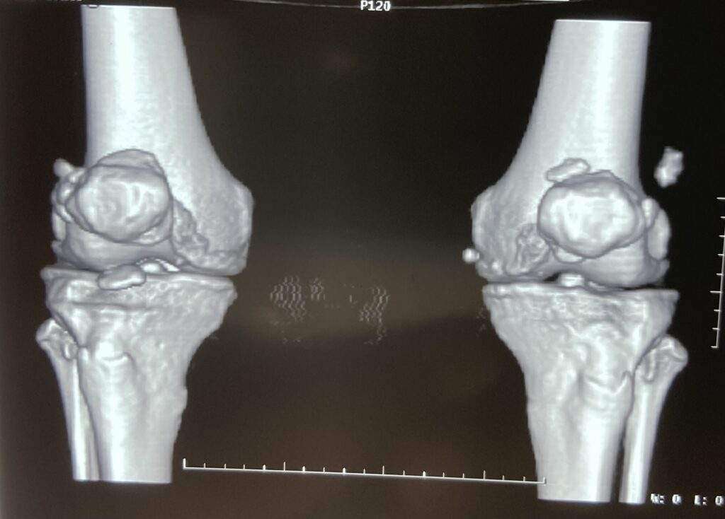
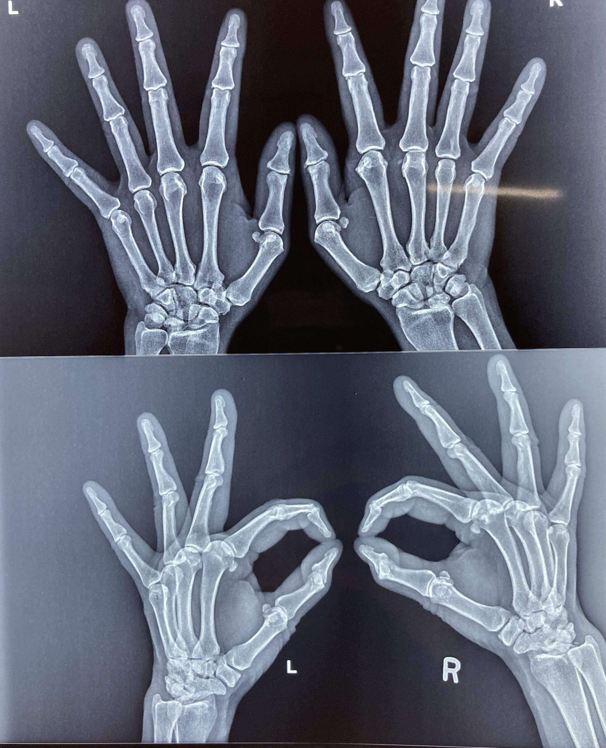
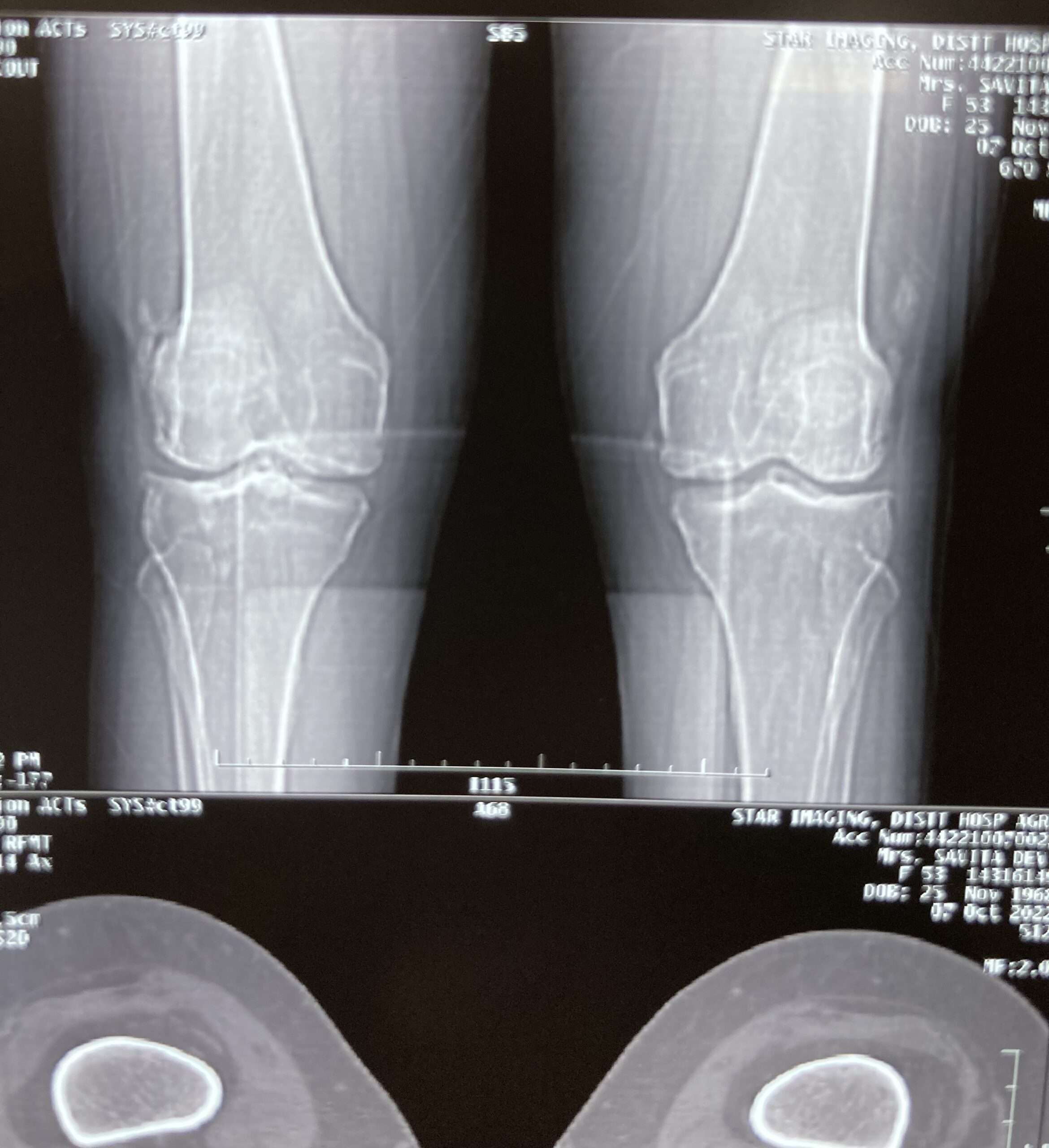
World Arthritis Day

World Arthritis Day 12 Oct

World Arthritis Day

World Arthritis Day 12 October 2022 – Theme, Importance and History

World Arthritis day is a global health awareness event organized every year on 12 October to create awareness about the rheumatic and musculoskeletal diseases, its impact on one’s life and educating people the symptoms & preventive measures and guiding for the early diagnosis to cope up any further complications. The day focuses on bringing people around the world on a single platform to raise voice to create enough opportunity to support and provide better treatment options to the people affected with rheumatic and musculoskeletal diseases (RMDs).

Importance of World Arthritis Day (WAD)

Arthritis is an inflammatory joint disorder, which affects the joints tissues around the joint, and other connective tissues, causing joint pain and stiffness. More than 100 types of arthritis exists, but most common are osteoarthritis and rheumatoid arthritis. Due to the lack of awareness and support, arthritis and its related condition has crippled much life around the world.

There is no specific treatment for the arthritis, treatment option varies based on the types, so it is imperative to understand the sign & symptoms and get early diagnosis to avail appropriate treatment. World Arthritis day (WAD), plays crucial role in encouraging people, concern medical fraternity and government across the world to participate in the awareness campaign to create better opportunity for the affected one.

World Arthritis Day 2022 Theme

This year 2022, the World Arthritis Day Theme is “It’s in your hands, take action“, addressing the world population to join hands for this cause to make this event fruitful for those deprived of the support.

The theme “It’s in your hands, take action” aims to encourage people with arthritis, their caregivers, families, and the general public to avail every opportunity to take action to improve their lifestyle.

World Arthritis Day History

World arthritis day (WAD) was established by Arthritis and Rheumatism International (ARI) and the first event was observed on 12 October 1996. Since then many global communities like Arthritis Foundation has joined the cause to raise awareness to fight the awareness gap, provide support and access to the communities, advocating for strong policies and supporting the research work. 

People living with Rheumatic and Musculoskeletal Diseases

Too many remain undiagnosed in the EU alone with an RMD. With an estimated one-hundred million currently undiagnosed and trying to cope with symptoms that are often overlooked – and frequently misdiagnosed. A large part of the world’s population is affected by diseases that impact their quality of life and participation in society – including access to the world of work. An inability to work reduces the self-esteem of the individual – and increases their dependency on state welfare, the healthcare system and their family and friends. People living with an RMD are therefore left unaware of their symptoms, opportunities for diagnosis and treatment, as well as the way to develop and execute a career plan to achieve independence.

Symptoms of Arthritis

  • Joint pain.
  • Stiffness and swelling in joints.
  • Your range of motion may also decrease.
  • Redness of the skin around the joint.
  • Unexplained joint pain usually accompanied by fever.
  • Difficulty in carrying out daily chores due to joint pain.
  • Pain killers also cannot cure joint pains.

Self-care tips to manage Joint Pain due to arthritis

  • Include a well-balanced diet with plenty of anti-inflammatory food such as green leafy vegetables, berries, ginger, nuts, legumes, and fibre.
  • Prioritize vitamin D and Calcium intake.
  • Maintain Your Ideal Weight as carrying excess body weight adds stress to our joints, especially the weight-bearing joints.
  • Practice regular physical activity followed by Low-Impact Exercises, which would put less stress on joints. Some of the Low-Impact Exercises that can be practice on daily basis are swimming, walking and cycling.
  • Engage in yoga and meditation to help your muscles relax.
  • Stop Smoking.
Dupuytrens Contracture

Dupuytrens Contracture

Dupuytrens Contracture

Overview

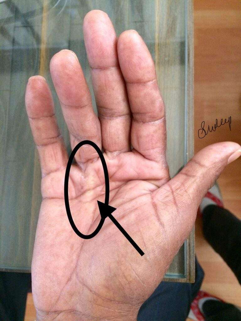
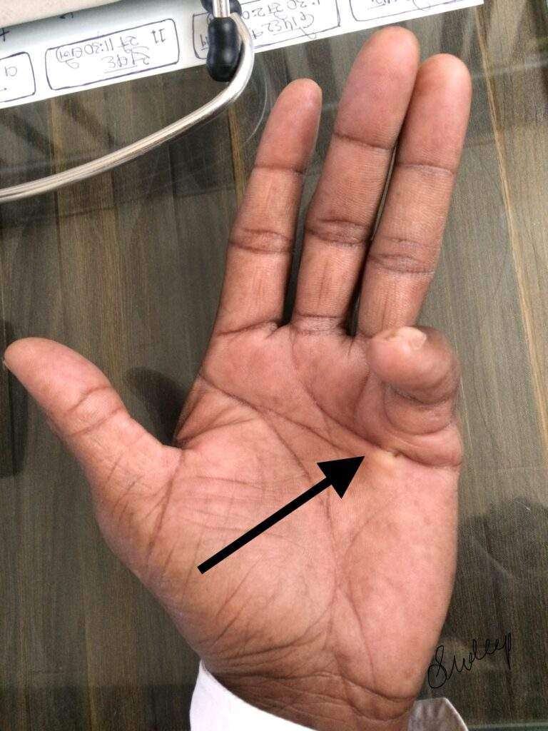
Dupuytrens (du-pwe-TRANZ) contracture is a hand deformity that usually develops over years. The condition affects a layer of tissue that lies under the skin of your palm. Knots of tissue form under the skin — eventually creating a thick cord that can pull one or more fingers into a bent position.

The affected fingers can’t be straightened completely, which can complicate everyday activities such as placing your hands in your pockets, putting on gloves or shaking hands.

Dupuytrens contracture mainly affects the two fingers farthest from the thumb, and occurs most often in older men of Northern European descent. A number of treatments are available to slow the progression of Dupuytrens contracture and relieve symptoms.

Symptoms

Dupuytrens contracture typically progresses slowly, over years. The condition usually begins as a thickening of the skin on the palm of your hand. As it progresses, the skin on your palm might appear puckered or dimpled. A firm lump of tissue can form on your palm. This lump might be sensitive to the touch but usually isn’t painful.

In later stages of Dupuytrens contracture, cords of tissue form under the skin on your palm and can extend up to your fingers. As these cords tighten, your fingers might be pulled toward your palm, sometimes severely.

The two fingers farthest from the thumb are most commonly affected, though the middle finger also can be involved. Only rarely are the thumb and index finger affected. Dupuytrens contracture can occur in both hands, though one hand is usually affected more severely.

Causes

Doctors don’t know what causes Dupuytrens contracture. There’s no evidence that hand injuries or occupations that involve vibrations to the hands cause the condition.

Risk factors

A number of factors are believed to increase your risk of the disease, including:

  • Age. Dupuytrens contracture occurs most commonly after the age of 50.
  • Sex. Men are more likely to develop Dupuytrens and to have more severe contractures than are women.
  • Ancestry. People of Northern European descent are at higher risk of the disease.
  • Family history. Dupuytrens contracture often runs in families.
  • Tobacco and alcohol use. Smoking is associated with an increased risk of Dupuytrens contracture, perhaps because of microscopic changes within blood vessels caused by smoking. Alcohol intake also is associated with Dupuytrens.
  • Diabetes. People with diabetes are reported to have an increased risk of Dupuytrens contracture.

Complications

Dupuytren’s contracture can make it difficult to perform certain functions using your hand. Since the thumb and index finger aren’t usually affected, many people don’t have much inconvenience or disability with fine motor activities such as writing. But as Dupuytrens contracture progresses, it can limit your ability to fully open your hand, grasp large objects or to get your hand into narrow places.

Diagnosis

In most cases, doctors can diagnose Dupuytrens contracture by the look and feel of your hands. Other tests are rarely necessary.

Your doctor will compare your hands to each other and check for puckering on the skin of your palms. He or she will also press on parts of your hands and fingers to check for toughened knots or bands of tissue.

Your doctor also might check to see if you can put your hand flat on a tabletop or other flat surface. Not being able to fully flatten your fingers indicates you have Dupuytrens contracture.

Treatment

If the disease progresses slowly, causes no pain and has little impact on your ability to use your hands for everyday tasks, you might not need treatment. Instead, you can wait and see if Dupuytrens contracture progresses. You may wish to follow the progression with a tabletop test, which you can do on your own.

Treatment involves removing or breaking apart the cords that are pulling your fingers toward your palm. This can be done in several ways. The choice of procedure depends on the severity of your symptoms and other health problems you may have.

Needling

This technique uses a needle, inserted through your skin, to puncture and break the cord of tissue that’s contracting a finger. Contractures often recur but the procedure can be repeated.

The main advantages of the needling technique are that there is no incision, it can be done on several fingers at the same time, and usually very little physical therapy is needed afterward. The main disadvantage is that it can’t be used in some places in the finger because it could damage a nerve or tendon.

Enzyme injections

Injecting a type of enzyme into the taut cord in your palm can soften and weaken it — allowing your doctor to later manipulate your hand in an attempt to break the cord and straighten your fingers. The FDA has approved collagenase Clostridium histolyticum for this purpose. The advantages and disadvantages of the enzyme injection are similar to needling. Enzyme injections are not offered at all medical institutions.

Surgery

Another option for people with advanced disease, limited function and progressing disease is to surgically remove the tissue in your palm affected by the disease. The main advantage to surgery is that it results in a more complete and longer-lasting release than that provided by the needle or enzyme methods. The main disadvantages are that physical therapy is usually needed after surgery, and recovery can take longer.

In some severe cases, especially if surgery has failed to correct the problem, surgeons remove all the tissue likely to be affected by Dupuytren’s contracture, including the attached skin. In these cases a skin graft is needed to cover the open wound. This surgery is the most invasive option and has the longest recovery time. People usually require months of intensive physical therapy afterward.

Lifestyle and home remedies

If you have mild Dupuytrens contracture, you can protect your hands by:

  • Avoiding a tight grip on tools by building up the handles with pipe insulation or cushion tape
  • Using gloves with heavy padding during heavy grasping tasks

However, your condition may persist or worsen, despite these precautions.

Preparing for your appointment

While you might first bring your symptoms to the attention of your family doctor, he or she might refer you to a specialist doctor.

What you can do

Before your appointment, you might want to write a list that answers the following questions:

  • Do you have a family history of this problem?
  • What treatments have you tried? Did they help?
  • What medications and supplements do you take regularly?

What to expect from your doctor

Your doctor might ask some of the following questions:

  • When did your symptoms begin?
  • Have they been getting worse?
  • Is your hand painful?
  • How does the contracture interfere with your day-to-day tasks?