Bhavishya Clinic+

Author name: sukum

Breathe easy this diwali

BREATHE EASY THIS DIWALI 2022

Breathe easy this diwali

Measures to breathe easy this Diwali?

Breathe easy this Diwali which is arguably India’s biggest festival which is synonymous with lights, lamps, colors and of course, firecrackers. However, this is also the time when air quality deteriorates drastically. The air is chocked with various forms of pollutants with the problem compounded by pollution caused by the increased movement of vehicles.

Patients visiting to the hospitals with complaints of severe respiratory disorders during Diwali festival have been increasing for the last five years. These include asthma, ARDS, and exacerbations of chronic lung disorders (COPD), all associated with breathing in polluted and toxic air. The problem of pollution is because of the effects of fireworks in Diwali. It is more severe in cities as the growing number of vehicles adds to the problem.

Air quality takes a deep plunge as the problem gets compounded. Depending on the size of these particles, they cause a variety of illnesses ranging from mild temporary irritation to serious chronic diseases such as cancer.

Particles with a diameter of 10 micrometers (PM 10) and less are considered the most harmful as they are small enough to evade the body’s filters and pass through the entire respiratory system. Travelling from the upper respiratory tract which consists of the nose, pharynx and larynx, the smaller particles end up accumulating in the lower respiratory tract (bronchioles, alveolar ducts, and alveolar sacs) where gas exchanges occur. These are eventually removed by the macrophages of the immune system.

When there is an increased number of particulate matter in the air along with gases, the chances of COPD exacerbations increase. Certain groups of people are particularly susceptible, such as:

Children – Children spend a lot of time playing outdoors and this is the reason why they run a high risk of getting affected by smog. Regular exposure to air pollution (smog) can cause asthma and even other harmful respiratory ailments.

People who do outdoor activities – Anyone who works outdoor is more susceptible to air pollution. Regular exposure and can deteriorate their health causing several respiratory diseases.

People with Asthma & Respiratory disease – People with asthma or chronic respiratory diseases are at a high risk of health hazards caused by smog. They will experience the adverse effects of air pollution much sooner compared to others. Continuous exposure to smog can aggravate lung diseases, heart disease and stroke.

Elderly People – Senior citizens have a weak immune system. Owing to this condition, elderly people are at increased risk of getting affected by air pollution, which can cause cardiac arrhythmias and heart attacks, asthma attacks and many more.

Dry hacking coughs, shortness of breath, chest tightness, wheezing, nasal complaint along with sneezing are some of the common complaints of patients during this time. Air pollution not only affects your lungs but other organs. Thus, it is important to know the health hazards during Diwali time and take precautions accordingly.

  1. Upper Respiratory Tract Infection: The Post-Diwali period usually witnesses a surge in the number of people with complaints of irritation in the eyes and throat, dry cough and fever. While last year’s smog lingered in the atmosphere, many people who otherwise remain healthy, also experienced irritation in eyes and chest and a general feeling of suffocation.
  2. Bronchitis: The deadly chemicals used in crackers release fumes and gases that can lead to the inflammation of the bronchial tubes leading to acute attacks.
  3. Asthma: The condition of asthma patients usually exacerbates during this period because of the toxic fumes in the air.
  4. COPD: The risk of Chronic Obstructive Pulmonary Disease (COPD) increases during festivals like Diwali when the level of suspended particulate matter becomes high in the air. It is characterized by prolonged cough with excess sputum.

Persons suffering from asthma, bronchitis and other respiratory ailments should start taking precautions several days before the festive season. Also, it is advisable for affected populations to avoid outdoor physical activities and wait until the levels of fine particles decrease. People suffering from Asthma, if it cannot be avoided should use a N95 face mask, which filters out at least 95% of airborne particles.

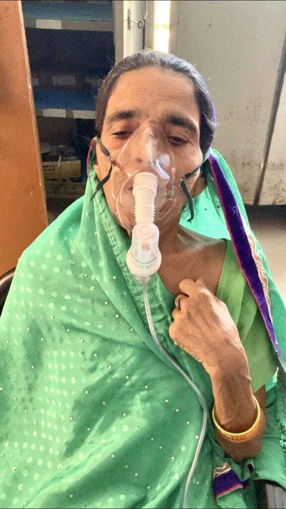
A normal surgical mask is of no use as pollutants can pass through it. It is important to keep inhalers handy. If one experiences breathlessness then it is advisable to move away from smoggy surroundings and sit in a room with the doors and windows shut and the air-conditioning on. Patients are supposed to continue their treatment for chronic lung disease regularly and consult pulmonologist at regular intervals. It is important to keep an inhaler or nebulizer at hand to dilate your airways. Rush to a hospital if you don’t feel better after inhalation.

Do’s & Don’ts to Stay Safe and breathe easy this Diwali

DOs

  • Use Multi Layered Masks: While going out, ensure that your nose and mouth are covered with a good quality mask to ban the entry of harmful particles in your respiratory tract. If you don’t have a mask, simply use a handkerchief or cloth to cover up.
  • Use Air Purifiers: Air inside closed spaces is more polluted than outside air. It’s advisable to install air purifiers in at homes and offices that allow only a little movement of air.
  • Use Air Purifying Plants: Your home and office must have plenty of plants that purify the air. A noteworthy point here is that there must be a variety of such plants and they must be spaced appropriately.
  • Use Air Quality Checking Apps: Before you go out, check the level of pollution through various Apps. If the air quality is severe, avoid going out at all.
  • Moisturize Your Nostrils: Moisturizing your nostrils helps in restricting the entry of fine particulate matter through your nose.

DON’Ts

  • Morning and Evening Walks/Runs: Air quality is worst in the mornings. Simply avoid morning and evening walks or running outside altogether till air quality improves.
  • Heavy Exercising: Do only light exercises, as heavy exercises require heavy breathing.
  • Eating Fried/Junk Food: Ensure you eat light, healthy, fiber rich and fat free food to feel healthier and fresh.
  • Travelling on two wheelers: Say No to travelling on two wheelers- this can be hazardous for your health.

Human life is above all and good health is a prerequisite of a happy life. We urge everyone to celebrate Diwali responsibly this year- in a way which makes every face glow with a smile and does not put anyone at any kind of health risk.

“Wishing You All, Happy & Safe Diwali”
calorie calculator

Calculate Your Optimal Calories in 2022

Mifflin – St. Jeor Calorie Calculator

Calculate Your Optimal Calories
Imperial
Metric
Basic Information
Activity Level
Your results:
Target calorie intake per day:
0

How many calories do I need?

An acceptable macronutrient distribution range for carbohydrates (45-65% of energy), protein (10-35% of energy), and fat (20-35% of energy; limit saturated and trans fats). These recommendations are broad and meant to cover the needs of many different people with different dietary situations. This is a much-discussed and debated issue, and the current understanding is that there is no one-size-fits-all recommendation. How many calories you consume and what percent of your calories come from each macronutrient can be manipulated to meet your individual needs and goals. However, consuming adequate protein at or above the recommended amount is necessary to maintain lean body mass.

About

The Mifflin-St Jeor equation is a widely used tool to determine the resting metabolic rate [RMR], which is defined as the number of calories burned while the body is in complete rest. RMR is also known as resting energy expenditure [REE]. The equation was developed by MD Mifflin and ST St Jeor and first introduced in a paper published in 1990. We are using this calorie calculator at our Bhavishya Clinic+

There are several equations for measuring RMR, including the most popular Harris-Benedict equation which was developed in 1919 and revised for accuracy in 1984. A comparative study of four predictive equations found that the Mifflin-St Jeor equation is more likely than the other equations to predict RMR to within 10% of that measured.

Formula

Females: (10*weight [kg]) + (6.25*height [cm]) – (5*age [years]) – 161
Males: (10*weight [kg]) + (6.25*height [cm]) – (5*age [years]) + 5

Multiply by scale factor for activity level:
Sedentary *1.2
Lightly active *1.375
Moderately active *1.55
Active *1.725
Very active *1.9

Basal metabolic rate is the amount of energy needed for your body’s physiological functions at rest, such as your breathing, your heart beating, and your brain activity.

This amount of energy required is measured in kilocalories, or often just denoted as calories. Therefore, BMR is your body’s calorie needs at rest with no extra activity.

In order to measure your exact BMR, you need to be in a lab setting and use indirect calorimetry. This measurement is taken under tightly controlled conditions and not easily accessible to most people.

Caloric expenditure measured by indirect calorimetry can then be added to physical activity energy expenditure to calculate total daily energy expenditure.

What is resting metabolic rate (RMR)?

Resting metabolic rate is the total number of calories burned when your body is completely at rest. RMR supports breathing, circulating blood, organ functions, and basic neurological functions. It is proportional to lean body mass and decreases approximately 0.01 kcal/min for each 1% increase in body fatness.

Hunger Vs Appetite

Lastly, take the time to understand some basic differences between hunger and appetite which are outlined below:

Hunger

It is considered a biological response to replenish the body’s energy reserves.

  • Protects us from starvation.
  • Usually triggered by an event occurring below the neckline:
    • Low blood sugar.
    • Empty (growling) stomach.
    • Hormone fluctuations.
    • Need to warm body (hypothermia).
  • Gradual onset, appearing after several hours without food and typically diminishes after eating.
  • It is generally satisfied by almost any food that provides energy (calories).

Appetite

It is considered a desire or interest to eat a specific food.

  • Usually triggered by an event occurring above the neckline consciously or subconsciously:
    • Thoughts, emotions and moods.
    • Social (e.g., happy hour)
    • Cultural (e.g., family)
    • Environmental (e.g., walking into a bakery).
  • More rapid onset and often independent of hunger.
  • Not time-dependent and may persist after eating.
  • Usually only satisfied by a specific food (e.g., sweet, salty) which may then evoke emotions and thoughts afterwards (e.g., pleasure, guilt, shame).

While RMR is an important component of TDEE, an accurate measurement remains elusive for many. Subsequently, we resort to mathematical formulas, but considering their potential errors, the values determined should always be considered a general estimate rather than an accurate value. Given this, there may also be value in including other methods as a guide to avoiding starvation. 

Lastly, while we need to acknowledge the fact that RMR is not entirely controllable, there are some influencing factors we can manipulate and should leverage every opportunity to exploit them.

Cataract

Cataract

Cataract
Cataract
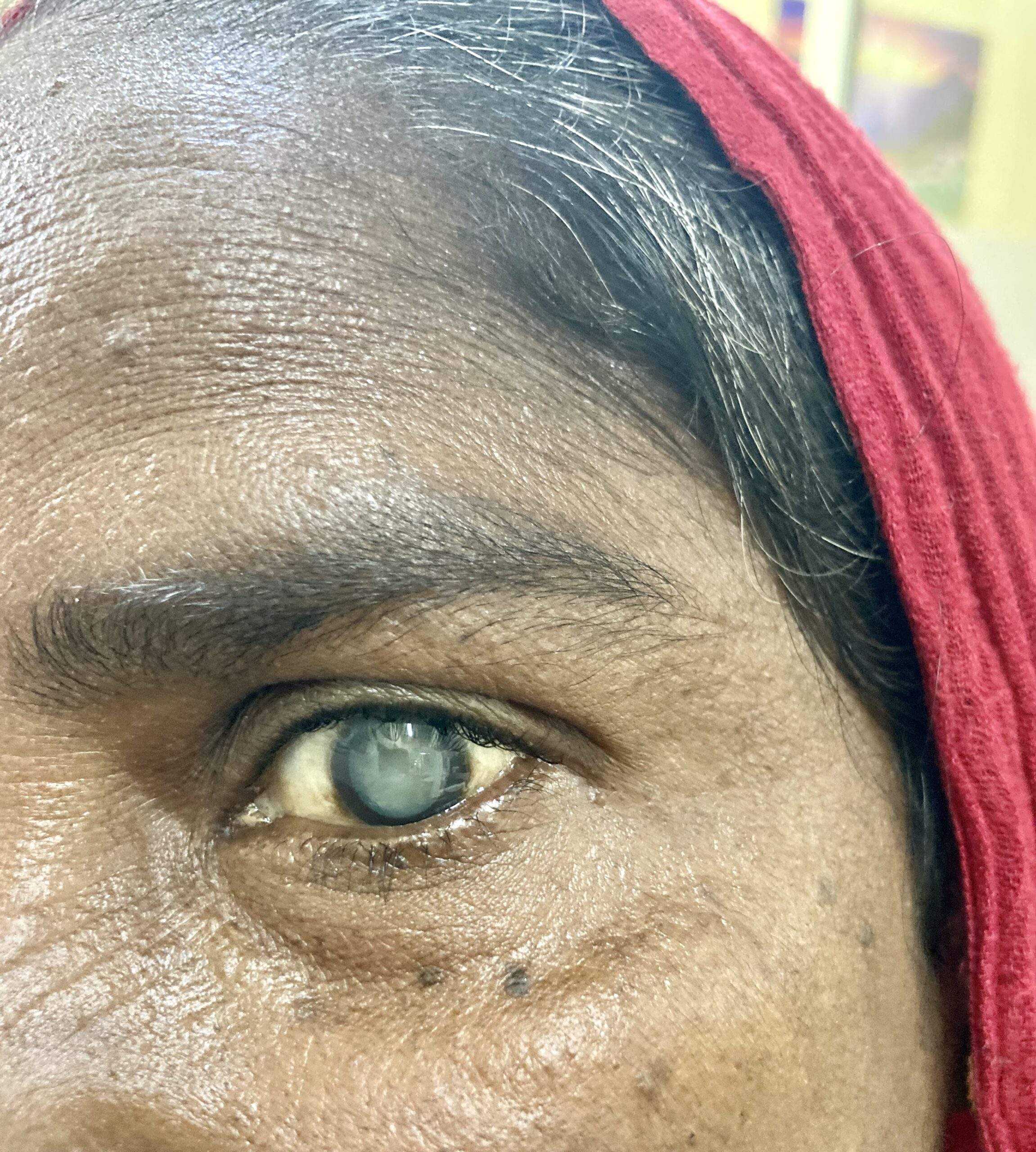
Matyre Cataract
Mature Cataract

Overview

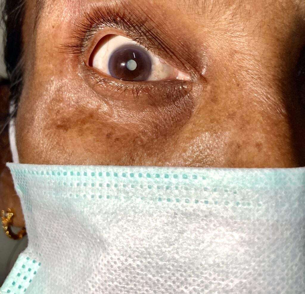
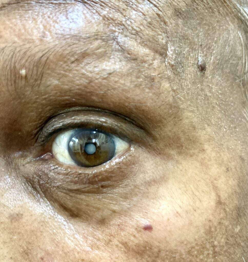
A cataract is a clouding of the normally clear lens of the eye. For people who have cataracts, seeing through cloudy lenses is a bit like looking through a frosty or fogged-up window. Clouded vision caused by cataracts can make it more difficult to read, drive a car (especially at night) or see the expression on a friend’s face.

Most cataracts develop slowly and don’t disturb your eyesight early on. But with time, cataracts will eventually interfere with your vision.

At first, stronger lighting and eyeglasses can help you deal with cataracts. But if impaired vision interferes with your usual activities, you might need cataract surgery. Fortunately, cataract surgery is generally a safe, effective procedure.

Symptoms

Signs and symptoms of cataracts include:

  • Clouded, blurred or dim vision
  • Increasing difficulty with vision at night
  • Sensitivity to light and glare
  • Need for brighter light for reading and other activities
  • Seeing “halos” around lights
  • Frequent changes in eyeglass or contact lens prescription
  • Fading or yellowing of colors
  • Double vision in a single eye
Cataract
Cataract in 62 year old OPD Patient

At first, the cloudiness in your vision caused by a cataract may affect only a small part of the eye’s lens and you may be unaware of any vision loss. As the cataract grows larger, it clouds more of your lens and distorts the light passing through the lens. This may lead to more-noticeable symptoms.

When to see a doctor

Make an appointment for an eye exam if you notice any changes in your vision. If you develop sudden vision changes, such as double vision or flashes of light, sudden eye pain, or sudden headache, see your doctor right away.

Causes

Most cataracts develop when aging or injury changes the tissue that makes up the eye’s lens. Proteins and fibers in the lens begin to break down, causing vision to become hazy or cloudy.

Some inherited genetic disorders that cause other health problems can increase your risk of cataracts. Cataracts can also be caused by other eye conditions, past eye surgery or medical conditions such as diabetes. Long-term use of steroid medications, too, can cause cataracts to develop.

How a cataract forms

A cataract is a cloudy lens. The lens is positioned behind the colored part of your eye (iris). The lens focuses light that passes into your eye, producing clear, sharp images on the retina — the light-sensitive membrane in the eye that functions like the film in a camera.

As you age, the lenses in your eyes become less flexible, less transparent and thicker. Age-related and other medical conditions cause proteins and fibers within the lenses to break down and clump together, clouding the lenses.

As the cataract continues to develop, the clouding becomes denser. A cataract scatters and blocks the light as it passes through the lens, preventing a sharply defined image from reaching your retina. As a result, your vision becomes blurred.

Cataracts generally develop in both eyes, but not always at the same rate. The cataract in one eye may be more advanced than the other, causing a difference in vision between eyes.

Types of cataracts

Cataract types include:

  • Cataracts affecting the center of the lens (nuclear cataracts). A nuclear cataract may at first cause more nearsightedness or even a temporary improvement in your reading vision. But with time, the lens gradually turns more densely yellow and further clouds your vision.As the cataract slowly progresses, the lens may even turn brown. Advanced yellowing or browning of the lens can lead to difficulty distinguishing between shades of color.
  • Cataracts that affect the edges of the lens (cortical cataracts). A cortical cataract begins as whitish, wedge-shaped opacities or streaks on the outer edge of the lens cortex. As it slowly progresses, the streaks extend to the center and interfere with light passing through the center of the lens.
  • Cataracts that affect the back of the lens (posterior subcapsular cataracts). A posterior subcapsular cataract starts as a small, opaque area that usually forms near the back of the lens, right in the path of light. A posterior subcapsular cataract often interferes with your reading vision, reduces your vision in bright light, and causes glare or halos around lights at night. These types of cataracts tend to progress faster than other types do.
  • Cataracts you’re born with (congenital cataracts). Some people are born with cataracts or develop them during childhood. These cataracts may be genetic, or associated with an intrauterine infection or trauma.These cataracts may also be due to certain conditions, such as myotonic dystrophy, galactosemia, neurofibromatosis type 2 or rubella. Congenital cataracts don’t always affect vision, but if they do, they’re usually removed soon after detection.

Risk factors

Factors that increase your risk of cataracts include:

  • Increasing age
  • Diabetes
  • Excessive exposure to sunlight
  • Smoking
  • Obesity
  • High blood pressure
  • Previous eye injury or inflammation
  • Previous eye surgery
  • Prolonged use of corticosteroid medications
  • Drinking excessive amounts of alcohol

Prevention

No studies have proved how to prevent cataracts or slow the progression of cataracts. But doctors think several strategies may be helpful, including:

  • Have regular eye examinations. Eye examinations can help detect cataracts and other eye problems at their earliest stages. Ask your doctor how often you should have an eye examination.
  • Quit smoking. Ask your doctor for suggestions about how to stop smoking. Medications, counseling and other strategies are available to help you.
  • Manage other health problems. Follow your treatment plan if you have diabetes or other medical conditions that can increase your risk of cataracts.
  • Choose a healthy diet that includes plenty of fruits and vegetables. Adding a variety of colorful fruits and vegetables to your diet ensures that you’re getting many vitamins and nutrients. Fruits and vegetables have many antioxidants, which help maintain the health of your eyes.Studies haven’t proved that antioxidants in pill form can prevent cataracts. But a large population study recently showed that a healthy diet rich in vitamins and minerals was associated with a reduced risk of developing cataracts. Fruits and vegetables have many proven health benefits and are a safe way to increase the amount of minerals and vitamins in your diet.
  • Wear sunglasses. Ultraviolet light from the sun may contribute to the development of cataracts. Wear sunglasses that block ultraviolet B (UVB) rays when you’re outdoors.
  • Reduce alcohol use. Excessive alcohol use can increase the risk of cataracts.

Diagnosis

To determine whether you have a cataract, your doctor will review your medical history and symptoms, and perform an eye examination. Your doctor may conduct several tests, including:

  • Visual acuity test. A visual acuity test uses an eye chart to measure how well you can read a series of letters. Your eyes are tested one at a time, while the other eye is covered. Using a chart or a viewing device with progressively smaller letters, your eye doctor determines if you have 20/20 vision or if your vision shows signs of impairment.
  • Slit-lamp examination. A slit lamp allows your eye doctor to see the structures at the front of your eye under magnification. The microscope is called a slit lamp because it uses an intense line of light, a slit, to illuminate your cornea, iris, lens, and the space between your iris and cornea. The slit allows your doctor to view these structures in small sections, which makes it easier to detect any tiny abnormalities.
  • Retinal exam. To prepare for a retinal exam, your eye doctor puts drops in your eyes to open your pupils wide (dilate). This makes it easier to examine the back of your eyes (retina). Using a slit lamp or a special device called an ophthalmoscope, your eye doctor can examine your lens for signs of a cataract.
  • Applanation tonometry. This test measures fluid pressure in your eye. There are multiple different devices available to do this.

Treatment

When your prescription glasses can’t clear your vision, the only effective treatment for cataracts is surgery.

When to consider cataract surgery

Talk with your eye doctor about whether surgery is right for you. Most eye doctors suggest considering cataract surgery when your cataracts begin to affect your quality of life or interfere with your ability to perform normal daily activities, such as reading or driving at night.

It’s up to you and your doctor to decide when cataract surgery is right for you. For most people, there is no rush to remove cataracts because they usually don’t harm the eyes. But cataracts can worsen faster in people with certain conditions, including diabetes, high blood pressure or obesity.

Delaying the procedure generally won’t affect how well your vision recovers if you later decide to have cataract surgery. Take time to consider the benefits and risks of cataract surgery with your doctor.

If you choose not to undergo cataract surgery now, your eye doctor may recommend periodic follow-up exams to see if your cataracts are progressing. How often you’ll see your eye doctor depends on your situation.

What happens during cataract surgery

Cataract surgery involves removing the clouded lens and replacing it with a clear artificial lens. The artificial lens, called an intraocular lens, is positioned in the same place as your natural lens. It remains a permanent part of your eye.

For some people, other eye problems prohibit the use of an artificial lens. In these situations, once the cataract is removed, vision may be corrected with eyeglasses or contact lenses.

Cataract surgery is generally done on an outpatient basis, which means you won’t need to stay in a hospital after the surgery. During cataract surgery, your eye doctor uses a local anesthetic to numb the area around your eye, but you usually stay awake during the procedure.

Cataract surgery is generally safe, but it carries a risk of infection and bleeding. Cataract surgery increases the risk of retinal detachment.

After the procedure, you’ll have some discomfort for a few days. Healing generally occurs within a few weeks.

If you need cataract surgery in both eyes, your doctor will schedule surgery to remove the cataract in the second eye after you’ve healed from the first surgery.

World Arthritis Day

World Arthritis Day 12 Oct

World Arthritis Day

World Arthritis Day 12 October 2022 – Theme, Importance and History

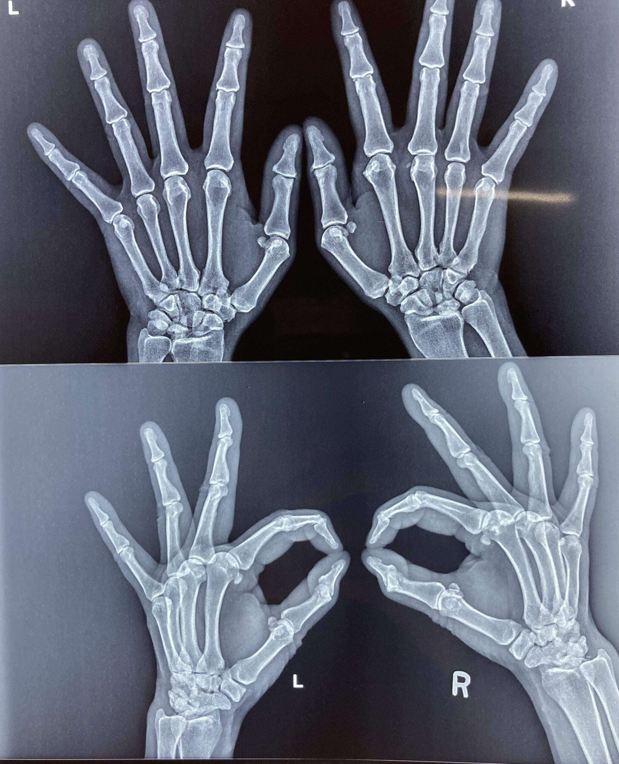
World Arthritis day is a global health awareness event organized every year on 12 October to create awareness about the rheumatic and musculoskeletal diseases, its impact on one’s life and educating people the symptoms & preventive measures and guiding for the early diagnosis to cope up any further complications. The day focuses on bringing people around the world on a single platform to raise voice to create enough opportunity to support and provide better treatment options to the people affected with rheumatic and musculoskeletal diseases (RMDs).

Importance of World Arthritis Day (WAD)

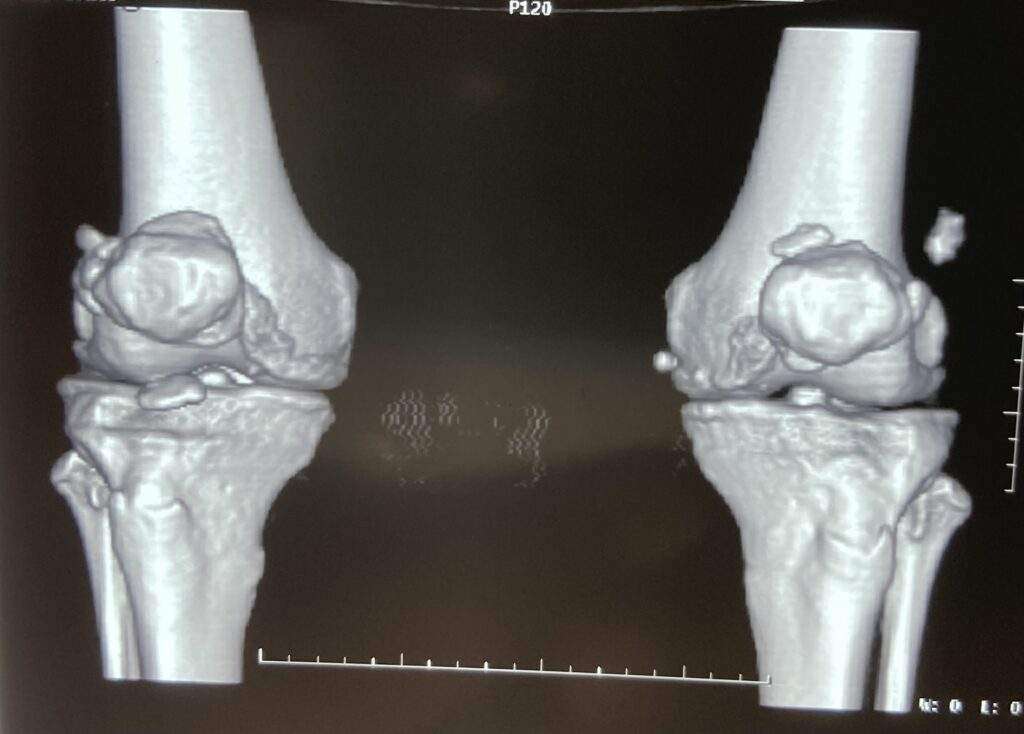
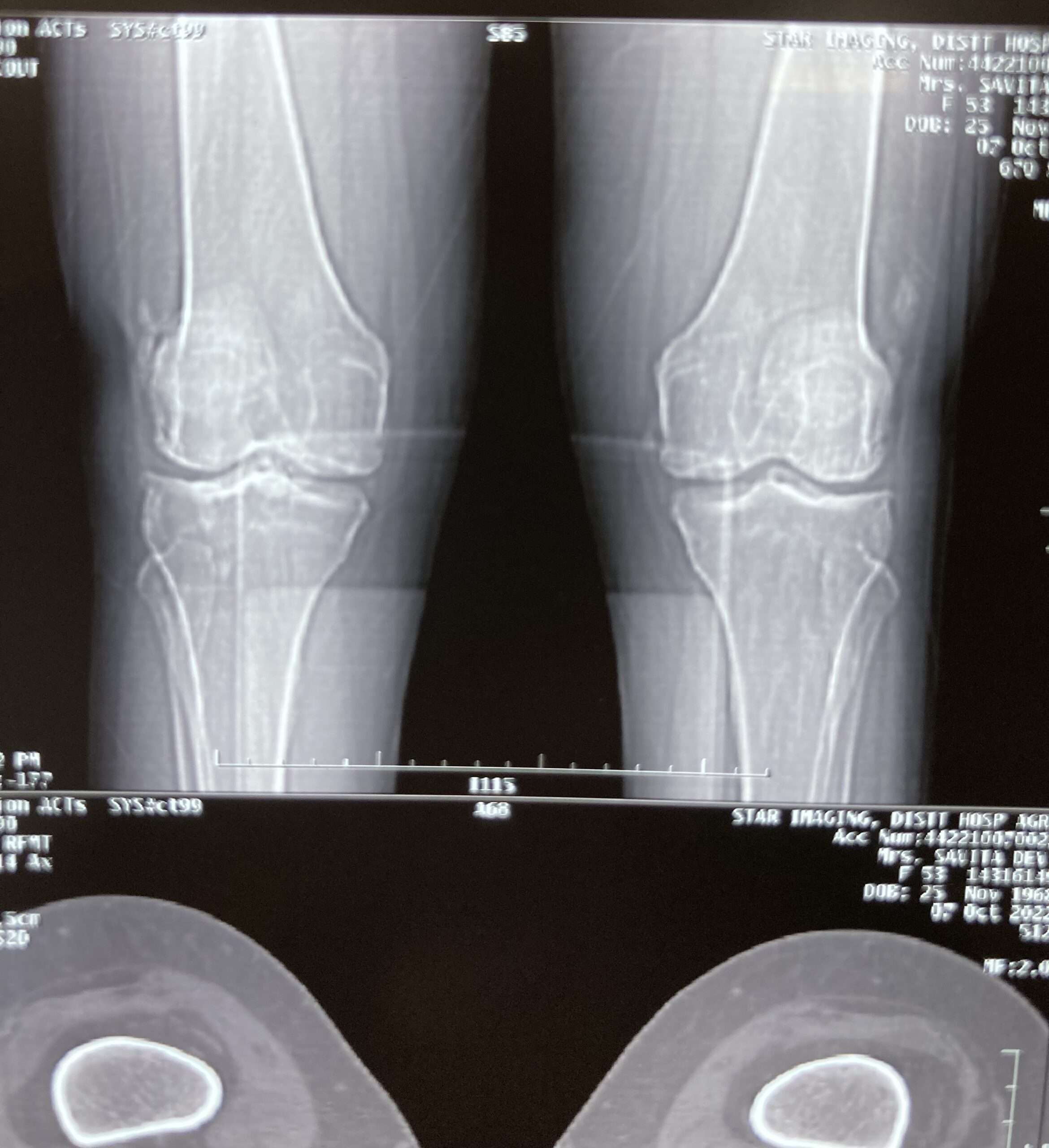
Arthritis is an inflammatory joint disorder, which affects the joints tissues around the joint, and other connective tissues, causing joint pain and stiffness. More than 100 types of arthritis exists, but most common are osteoarthritis and rheumatoid arthritis. Due to the lack of awareness and support, arthritis and its related condition has crippled much life around the world.

There is no specific treatment for the arthritis, treatment option varies based on the types, so it is imperative to understand the sign & symptoms and get early diagnosis to avail appropriate treatment. World Arthritis day (WAD), plays crucial role in encouraging people, concern medical fraternity and government across the world to participate in the awareness campaign to create better opportunity for the affected one.

World Arthritis Day 2022 Theme

This year 2022, the World Arthritis Day Theme is “It’s in your hands, take action“, addressing the world population to join hands for this cause to make this event fruitful for those deprived of the support.

The theme “It’s in your hands, take action” aims to encourage people with arthritis, their caregivers, families, and the general public to avail every opportunity to take action to improve their lifestyle.

World Arthritis Day History

World arthritis day (WAD) was established by Arthritis and Rheumatism International (ARI) and the first event was observed on 12 October 1996. Since then many global communities like Arthritis Foundation has joined the cause to raise awareness to fight the awareness gap, provide support and access to the communities, advocating for strong policies and supporting the research work. 

People living with Rheumatic and Musculoskeletal Diseases

Too many remain undiagnosed in the EU alone with an RMD. With an estimated one-hundred million currently undiagnosed and trying to cope with symptoms that are often overlooked – and frequently misdiagnosed. A large part of the world’s population is affected by diseases that impact their quality of life and participation in society – including access to the world of work. An inability to work reduces the self-esteem of the individual – and increases their dependency on state welfare, the healthcare system and their family and friends. People living with an RMD are therefore left unaware of their symptoms, opportunities for diagnosis and treatment, as well as the way to develop and execute a career plan to achieve independence.

Symptoms of Arthritis

  • Joint pain.
  • Stiffness and swelling in joints.
  • Your range of motion may also decrease.
  • Redness of the skin around the joint.
  • Unexplained joint pain usually accompanied by fever.
  • Difficulty in carrying out daily chores due to joint pain.
  • Pain killers also cannot cure joint pains.

Self-care tips to manage Joint Pain due to arthritis

  • Include a well-balanced diet with plenty of anti-inflammatory food such as green leafy vegetables, berries, ginger, nuts, legumes, and fibre.
  • Prioritize vitamin D and Calcium intake.
  • Maintain Your Ideal Weight as carrying excess body weight adds stress to our joints, especially the weight-bearing joints.
  • Practice regular physical activity followed by Low-Impact Exercises, which would put less stress on joints. Some of the Low-Impact Exercises that can be practice on daily basis are swimming, walking and cycling.
  • Engage in yoga and meditation to help your muscles relax.
  • Stop Smoking.
Dupuytrens Contracture

Dupuytrens Contracture

Dupuytrens Contracture

Overview

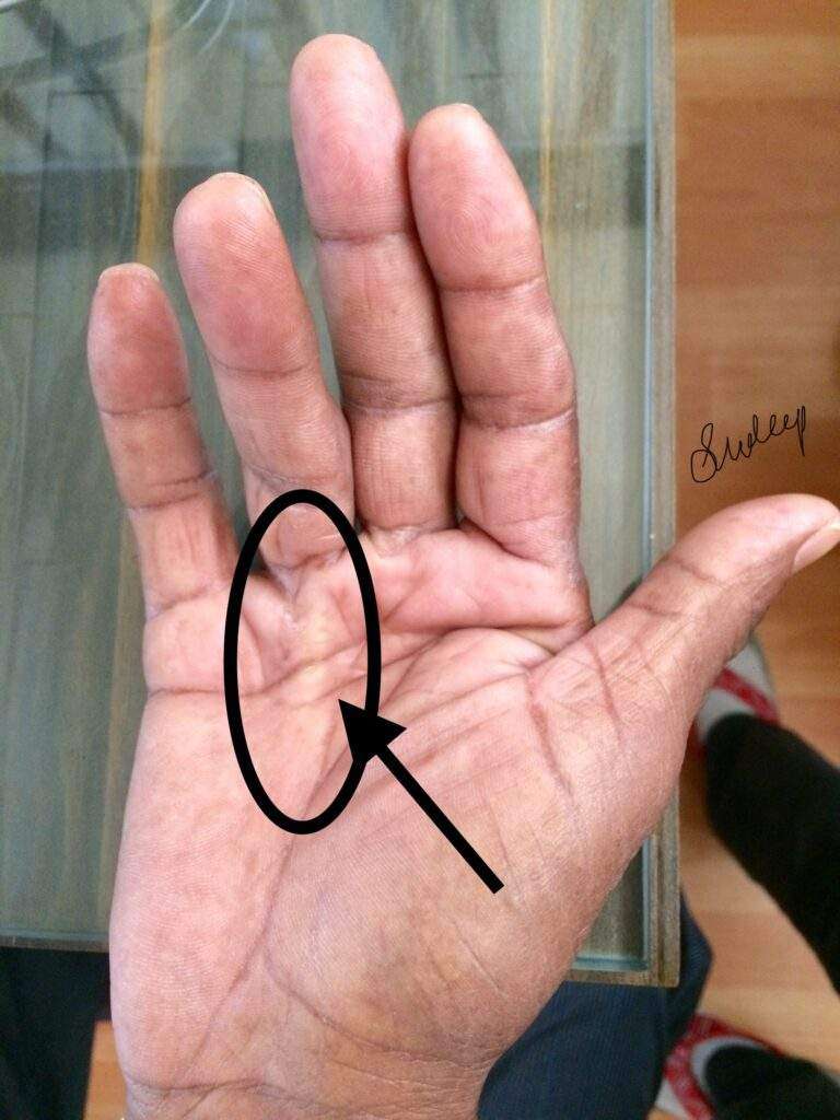
Dupuytrens (du-pwe-TRANZ) contracture is a hand deformity that usually develops over years. The condition affects a layer of tissue that lies under the skin of your palm. Knots of tissue form under the skin — eventually creating a thick cord that can pull one or more fingers into a bent position.

The affected fingers can’t be straightened completely, which can complicate everyday activities such as placing your hands in your pockets, putting on gloves or shaking hands.

Dupuytrens contracture mainly affects the two fingers farthest from the thumb, and occurs most often in older men of Northern European descent. A number of treatments are available to slow the progression of Dupuytrens contracture and relieve symptoms.

Symptoms

Dupuytrens contracture typically progresses slowly, over years. The condition usually begins as a thickening of the skin on the palm of your hand. As it progresses, the skin on your palm might appear puckered or dimpled. A firm lump of tissue can form on your palm. This lump might be sensitive to the touch but usually isn’t painful.

In later stages of Dupuytrens contracture, cords of tissue form under the skin on your palm and can extend up to your fingers. As these cords tighten, your fingers might be pulled toward your palm, sometimes severely.

The two fingers farthest from the thumb are most commonly affected, though the middle finger also can be involved. Only rarely are the thumb and index finger affected. Dupuytrens contracture can occur in both hands, though one hand is usually affected more severely.

Causes

Doctors don’t know what causes Dupuytrens contracture. There’s no evidence that hand injuries or occupations that involve vibrations to the hands cause the condition.

Risk factors

A number of factors are believed to increase your risk of the disease, including:

  • Age. Dupuytrens contracture occurs most commonly after the age of 50.
  • Sex. Men are more likely to develop Dupuytrens and to have more severe contractures than are women.
  • Ancestry. People of Northern European descent are at higher risk of the disease.
  • Family history. Dupuytrens contracture often runs in families.
  • Tobacco and alcohol use. Smoking is associated with an increased risk of Dupuytrens contracture, perhaps because of microscopic changes within blood vessels caused by smoking. Alcohol intake also is associated with Dupuytrens.
  • Diabetes. People with diabetes are reported to have an increased risk of Dupuytrens contracture.

Complications

Dupuytren’s contracture can make it difficult to perform certain functions using your hand. Since the thumb and index finger aren’t usually affected, many people don’t have much inconvenience or disability with fine motor activities such as writing. But as Dupuytrens contracture progresses, it can limit your ability to fully open your hand, grasp large objects or to get your hand into narrow places.

Diagnosis

In most cases, doctors can diagnose Dupuytrens contracture by the look and feel of your hands. Other tests are rarely necessary.

Your doctor will compare your hands to each other and check for puckering on the skin of your palms. He or she will also press on parts of your hands and fingers to check for toughened knots or bands of tissue.

Your doctor also might check to see if you can put your hand flat on a tabletop or other flat surface. Not being able to fully flatten your fingers indicates you have Dupuytrens contracture.

Treatment

If the disease progresses slowly, causes no pain and has little impact on your ability to use your hands for everyday tasks, you might not need treatment. Instead, you can wait and see if Dupuytrens contracture progresses. You may wish to follow the progression with a tabletop test, which you can do on your own.

Treatment involves removing or breaking apart the cords that are pulling your fingers toward your palm. This can be done in several ways. The choice of procedure depends on the severity of your symptoms and other health problems you may have.

Needling

This technique uses a needle, inserted through your skin, to puncture and break the cord of tissue that’s contracting a finger. Contractures often recur but the procedure can be repeated.

The main advantages of the needling technique are that there is no incision, it can be done on several fingers at the same time, and usually very little physical therapy is needed afterward. The main disadvantage is that it can’t be used in some places in the finger because it could damage a nerve or tendon.

Enzyme injections

Injecting a type of enzyme into the taut cord in your palm can soften and weaken it — allowing your doctor to later manipulate your hand in an attempt to break the cord and straighten your fingers. The FDA has approved collagenase Clostridium histolyticum for this purpose. The advantages and disadvantages of the enzyme injection are similar to needling. Enzyme injections are not offered at all medical institutions.

Surgery

Another option for people with advanced disease, limited function and progressing disease is to surgically remove the tissue in your palm affected by the disease. The main advantage to surgery is that it results in a more complete and longer-lasting release than that provided by the needle or enzyme methods. The main disadvantages are that physical therapy is usually needed after surgery, and recovery can take longer.

In some severe cases, especially if surgery has failed to correct the problem, surgeons remove all the tissue likely to be affected by Dupuytren’s contracture, including the attached skin. In these cases a skin graft is needed to cover the open wound. This surgery is the most invasive option and has the longest recovery time. People usually require months of intensive physical therapy afterward.

Lifestyle and home remedies

If you have mild Dupuytrens contracture, you can protect your hands by:

  • Avoiding a tight grip on tools by building up the handles with pipe insulation or cushion tape
  • Using gloves with heavy padding during heavy grasping tasks

However, your condition may persist or worsen, despite these precautions.

Preparing for your appointment

While you might first bring your symptoms to the attention of your family doctor, he or she might refer you to a specialist doctor.

What you can do

Before your appointment, you might want to write a list that answers the following questions:

  • Do you have a family history of this problem?
  • What treatments have you tried? Did they help?
  • What medications and supplements do you take regularly?

What to expect from your doctor

Your doctor might ask some of the following questions:

  • When did your symptoms begin?
  • Have they been getting worse?
  • Is your hand painful?
  • How does the contracture interfere with your day-to-day tasks?

Deep Vein Thrombosis

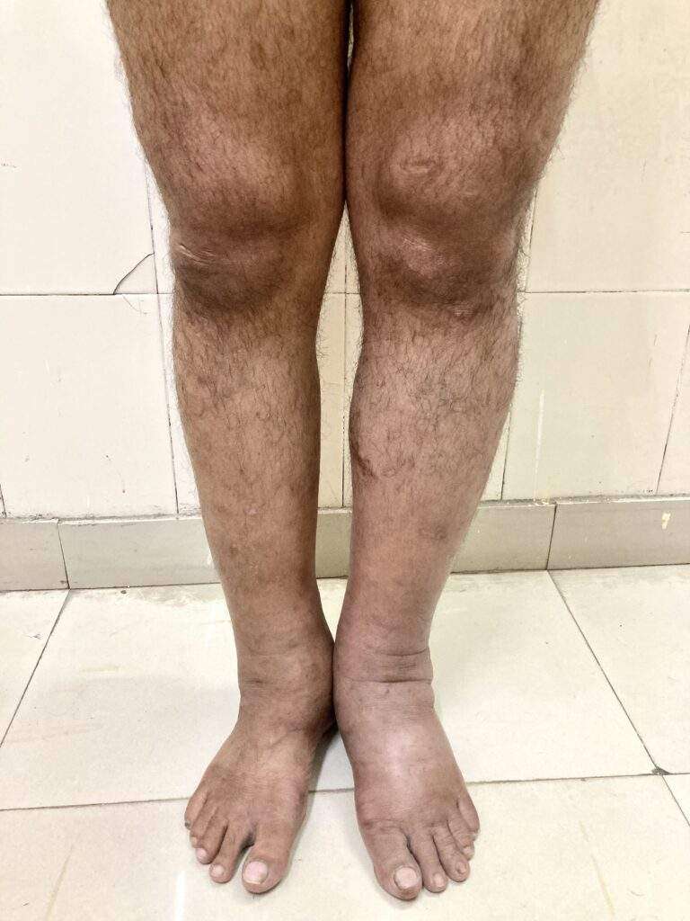
Blood clots are lifesavers when they seal a cut. They can be dangerous, even deadly, when they form inside an artery or vein. Deep vein thrombosis (sometimes called DVT) is the formation of a blood clot in a large leg vein. It can also occur in an arm vein. Deep vein thrombosis can lead to a pulmonary embolism, or sometimes a stroke.

Blood that circulates to the legs and feet must flow against gravity on its journey back to the heart. The trip is aided by the contraction of leg muscles during walking or fidgeting. The contractions squeeze veins, pushing blood through them. Small flaps, or valves, inside the veins keep blood flowing in the direction of the heart.

Anything that slows blood flow through the arms and legs can set the stage for a blood clot to form. This can range from having an arm or leg immobilized in a cast to prolonged sitting or being confined to bed. Things that make blood more likely to clot, such as genetic disorders and cancer, are other triggers for deep-vein thrombosis.

Symptoms of deep vein thrombosis and pulmonary embolism

Deep vein thrombosis can develop silently. It can also cause:

  • pain or tenderness in a leg or arm that gets worse with time, not better
  • swelling in one leg or arm
  • a reddish or bluish tinge to the skin of one leg or arm
  • a leg or arm that feels warm to the touch.

The symptoms of pulmonary embolism include:

  • difficulty breathing
  • chest pain or discomfort that worsens with a deep breath or cough
  • coughing up blood
  • a fast heart rate
  • sudden lightheadedness or fainting

Diagnosing deep vein thrombosis and pulmonary embolism

To diagnose DVT, your doctor will examine your legs to check for swelling and tenderness. He or she will ask about your symptoms and risk factors.

Based on the findings, your doctor may order a D-Dimer blood test or an ultrasound of your legs.

The blood test measures the level of a chemical called D-Dimer. It is almost always abnormally high when blood clots are actively forming in the body.

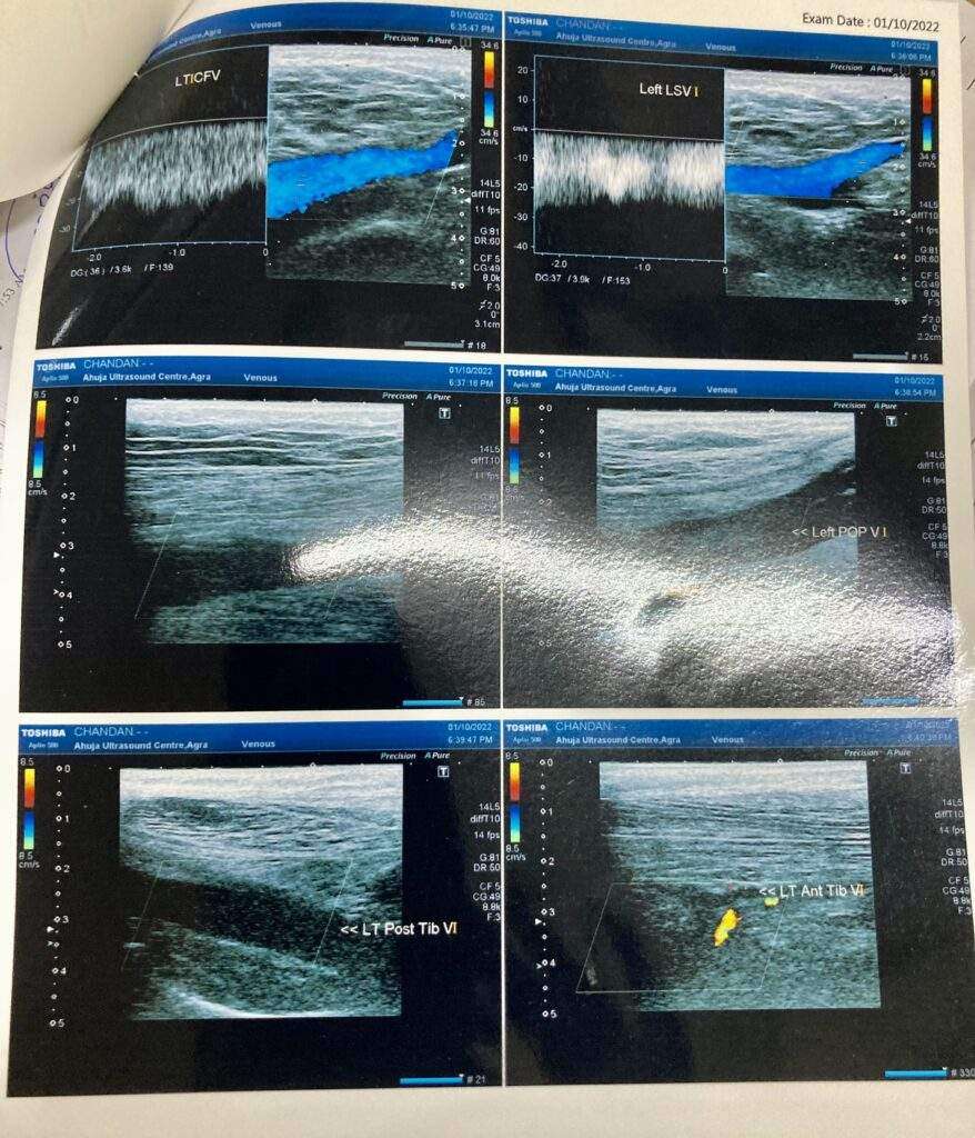
An ultrasound of your legs is done to look for blood flow problems in your veins. This procedure is called a lower extremity non-invasive test, or LENI. If the LENI shows evidence of a blood clot, your doctor will diagnose DVT.

If the initial LENI is negative, it does not mean that there is no clot. It may be too early to see the full effect of the clot. Your doctor may ask that you return in three to four days for a repeat LENI.

If your doctor suspects you have a pulmonary embolism, he or she will first try to determine if you have DVT. If the LENI shows one or more blood clots in your leg veins, and you have symptoms of a pulmonary embolism, an embolism is the most likely diagnosis.

Or your doctor may order computed tomography (CT) of the chest. The test requires an IV injection of dye to look for blood clots in the pulmonary arteries. People that have impaired kidney function or an allergy to the dye might need a different type of lung scan called a V/Q scan to examine lung blood flow.

Treating deep vein thrombosis

The initial treatment for a DVT or pulmonary embolism is heparin or one of the newer oral anti-coagulant drugs. These medications act on certain blood proteins to prevent new blood clot formation and therefore help unwanted clots get smaller. They are commonly called “blood thinners.”

There are two main types of heparin. The oldest type of heparin is best administered by a constant intravenous infusion. Another type of heparin is called low-molecular-weight heparin. It is injected under the skin once or twice per day.

Two of the newer anti-coagulant drugs are approved for initial treatment of DVT and pulmonary embolism: rivaroxaban (Xarelto) and apixaban (Eliquis).

If you have a DVT without a pulmonary embolism, you may not need to be hospitalized. You could be treated at home with injections of a low-molecular-weight heparin or either rivaroxaban or apixaban.

Some people may need to start therapy in the hospital. In this case, the type of heparin used is determined by many factors. These include body weight, kidney function and other circumstances.

If you have a pulmonary embolism, you will probably be hospitalized. If so, you likely will be treated with either type of heparin initially. But oral rivaroxaban or apixaban could be an option instead of heparin if your pulmonary embolism is small.

If you are started on either IV heparin or low-molecular weight heparin shots under the skin, your doctor will transition you to an oral drug. Traditional oral therapy has been warfarin (Coumadin). For decades, it was the only oral drug to treat DVT and pulmonary embolism.

In addition to rivaroxaban and apixaban, two other oral anti-coagulant drugs can be used after heparin: dabigatran (Pradaxa) and edoxapan (Savaysa). More of these types of drugs will be approved soon.

Warfarin takes a few days to start working. Once a blood test shows that warfarin is effective, you will stop taking heparin. You will continue taking warfarin for several months or longer.

During the first few weeks that you take warfarin, you will continue to need frequent blood tests to make sure you are taking the right amount. Once your blood test results consistently show that you are taking the right amount of medication, blood can be drawn every two to four weeks.

Some foods—especially green, leafy vegetables that contain large amounts of vitamin K—can alter the blood-thinning action of warfarin. Ask your doctor or pharmacist for a list of these foods. You can continue to eat these foods as long as you eat approximately the same amount of them each day. That way, the effect on your medication will be consistent.

Other medications can also affect how warfarin works in your body. Tell any doctor who is prescribing medications for you that you are taking warfarin.

The new novel oral anti-coagulants don’t require regular blood testing. They are given in a fixed dose. The other advantage is not worrying about eating food with too much vitamin K.

Enlarged Spleen

Enlarged Spleen (Splenomegaly)

Enlarged Spleen
Enlarged Spleen

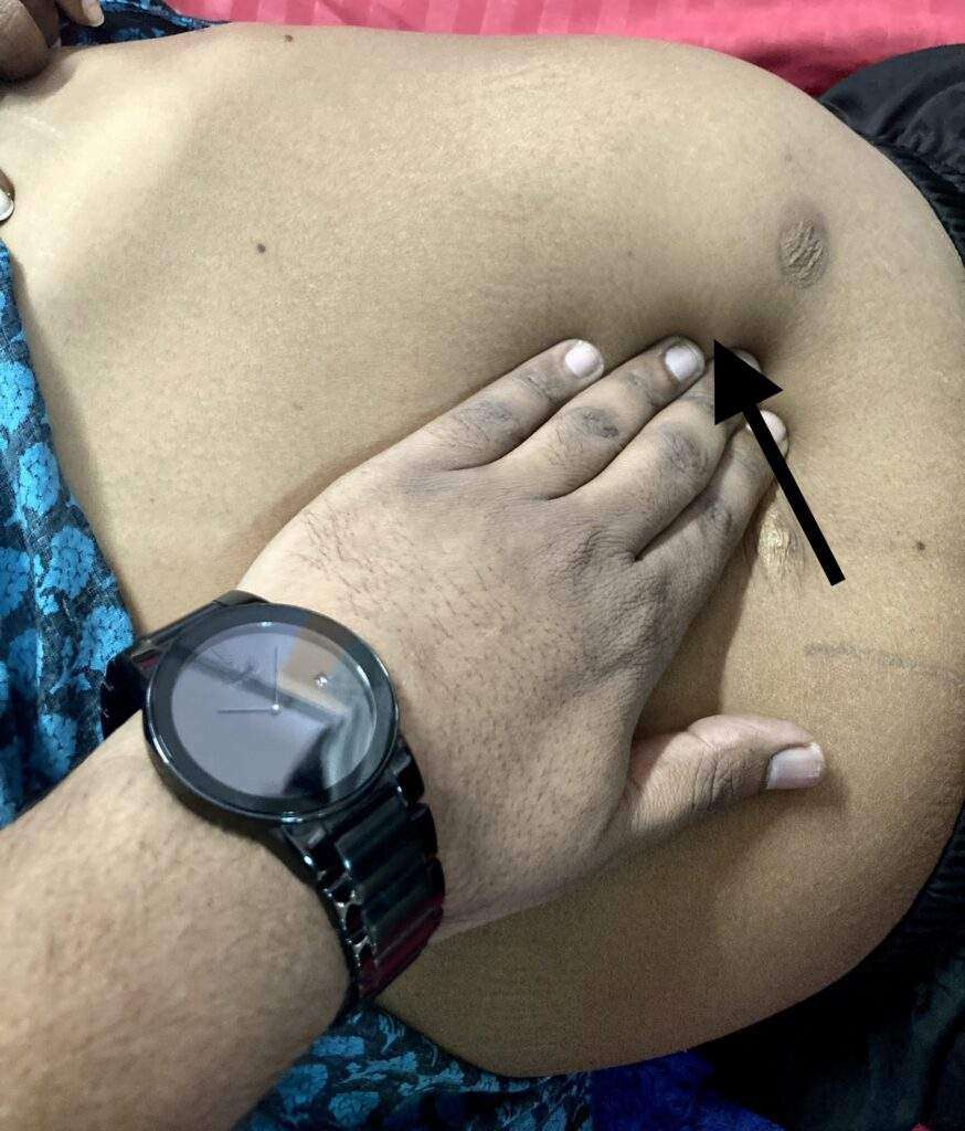
Rule of Odds in spleen: 1” thick, 3” broad, 5” long, 7” ounces weight, underlies 9-11 ribs

A normal, healthy spleen is up to 12 cm long and 70 g in weight. An enlarged spleen may be up to 20 cm long and can weigh more than 1,000 g. Several things can cause your spleen to enlarge, including inflammation, fat storage, pooled blood, benign or malignant growths and overproduction of cells. Some causes are temporary and others may indicate a chronic or progressive condition.

Symptoms

An enlarged spleen typically causes no signs or symptoms, but sometimes it causes:

  • Pain or fullness in the left upper belly that can spread to the left shoulder
  • A feeling of fullness without eating or after eating a small amount because the spleen is pressing on your stomach
  • Low red blood cells (anemia)
  • Frequent infections
  • Bleeding easily

When to see a doctor

See your doctor promptly if you have pain in your left upper belly, especially if it’s severe or the pain gets worse when you take a deep breath.

Causes

A number of infections and diseases can cause an enlarged spleen. The enlargement might be temporary, depending on treatment. Contributing factors include:

  • Viral infections, such as mononucleosis
  • Bacterial infections, such as syphilis or an infection of your heart’s inner lining (endocarditis)
  • Parasitic infections, such as malaria
  • Cirrhosis and other diseases affecting the liver
  • Various types of hemolytic anemia — a condition characterized by early destruction of red blood cells
  • Blood cancers, such as leukemia and myeloproliferative neoplasms, and lymphomas, such as Hodgkin’s disease
  • Metabolic disorders, such as Gaucher disease and Niemann-Pick disease
  • Pressure on the veins in the spleen or liver or a blood clot in these veins
  • Autoimmune conditions, such as lupus or sarcoidosis

How the spleen works

Your spleen is tucked below your rib cage next to your stomach on the left side of your belly. Its size generally relates to your height, weight and sex.

This soft, spongy organ performs several critical jobs, such as:

  • Filtering out and destroying old, damaged blood cells
  • Preventing infection by producing white blood cells (lymphocytes) and acting as a first line of defense against disease-causing organisms
  • Storing red blood cells and platelets, which help your blood clot

An enlarged spleen affects each of these jobs. When it’s enlarged, your spleen may not function as usual.

Risk factors

Anyone can develop an enlarged spleen at any age, but certain groups are at higher risk, including:

  • Children and young adults with infections, such as mononucleosis
  • People who have Gaucher disease, Niemann-Pick disease, and several other inherited metabolic disorders affecting the liver and spleen
  • People who live in or travel to areas where malaria is common

Complications

Potential complications of an enlarged spleen are:

  • Infection. An enlarged spleen can reduce the number of healthy red blood cells, platelets and white cells in your bloodstream, leading to more frequent infections. Anemia and increased bleeding also are possible.
  • Ruptured spleen. Even healthy spleens are soft and easily damaged, especially in car crashes. The possibility of rupture is much greater when your spleen is enlarged. A ruptured spleen can cause life-threatening bleeding in your belly.

Diagnosis

An enlarged spleen is usually detected during a physical exam. Your doctor can often feel it by gently examining your left upper belly. However, in some people — especially those who are slender — a healthy, normal-sized spleen can sometimes be felt during an exam.

Your doctor might order these tests to confirm the diagnosis of an enlarged spleen:

  • Blood tests, such as a complete blood count to check the number of red blood cells, white blood cells and platelets in your system and liver function
  • Ultrasound or CT scan to help determine the size of your spleen and whether it’s crowding other organs
  • MRI to trace blood flow through the spleen

Finding the cause

Sometimes more testing is needed to find the cause of an enlarged spleen, including a bone marrow biopsy exam.

A sample of solid bone marrow may be removed in a procedure called a bone marrow biopsy. Or you might have a bone marrow aspiration, which removes the liquid portion of your marrow. Both procedures might be done at the same time.

Liquid and solid bone marrow samples are usually taken from the pelvis. A needle is inserted into the bone through an incision. You’ll receive either a general or a local anesthetic before the test to ease discomfort.

A needle biopsy of the spleen is rare because of the risk of bleeding.

Your doctor might recommend surgery to remove your spleen (splenectomy) for diagnostic purposes when there’s no identifiable cause for the enlargement. More often, the spleen is removed as treatment. After surgery to remove it, the spleen is examined under a microscope to check for possible lymphoma of the spleen.

Treatment

Treatment for an enlarged spleen focuses on the what’s causing it. For example, if you have a bacterial infection, treatment will include antibiotics.

Watchful waiting

If you have an enlarged spleen but don’t have symptoms and the cause can’t be found, your doctor might suggest watchful waiting. You see your doctor for reevaluation in 6 to 12 months or sooner if you develop symptoms.

Spleen removal surgery

If an enlarged spleen causes serious complications or the cause can’t be identified or treated, surgery to remove your spleen (splenectomy) might be an option. In chronic or critical cases, surgery might offer the best hope for recovery.

Elective spleen removal requires careful consideration. You can live an active life without a spleen, but you’re more likely to get serious or even life-threatening infections after spleen removal.

Reducing infection risk after surgery

After spleen removal, certain steps can help reduce your risk of infection, including:

  • A series of vaccinations before and after the splenectomy. These include the pneumococcal (Pneumovax 23), meningococcal and haemophilus influenzae type b (Hib) vaccines, which protect against pneumonia, meningitis and infections of the blood, bones and joints. You’ll also need the pneumococcal vaccine every five years after surgery.
  • Taking penicillin or other antibiotics after your surgery and anytime you or your doctor suspects the possibility of an infection.
  • Calling your doctor at the first sign of a fever, which could indicate an infection.
  • Avoiding travel to parts of the world where certain diseases, such as malaria, are common.

A note from Bhavishya Clinic

An enlarged spleen is a symptom that healthcare providers need to investigate. Whether or not it’s causing you discomfort, it indicates an underlying condition that may need treatment. When it’s temporary, an enlarged spleen won’t harm your overall health. But chronic swelling could damage and endanger your spleen. Your healthcare provider will treat it by treating the underlying cause.

Down Syndrome

Down Syndrome I Trisomy of 21 Chromosome

Down Syndrome
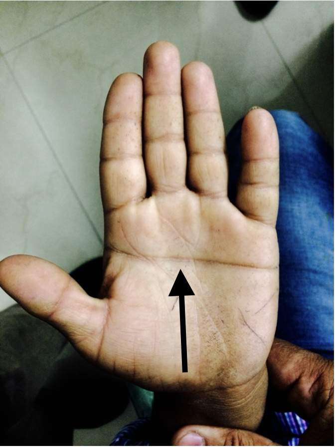
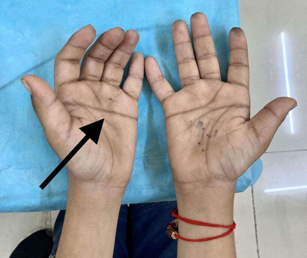
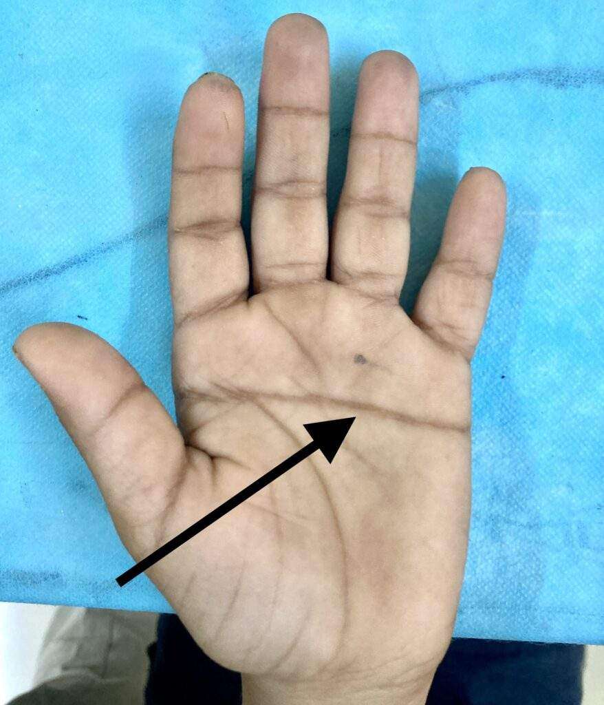
Simian Crease
Mongoloid facies with
Orbital Hypertelorism

What is Down Syndrome?

Down syndrome is a disorder caused by a problem with the chromosomes — the pieces of DNA that have the blueprint for the human body. Normally a person has two copies of each chromosome, but a person with Down syndrome has three copies of chromosome 21. The condition also is called trisomy 21.  

In a few cases, the extra copy is part of another chromosome (translocation), or found in only some of the person’s cells (mosaicism). 

The extra DNA makes the physical and mental characteristics, which include a small head that is flattened in the back; slanted eyes; extra skin folds at the corners of the eyes; small ears, nose and mouth; big-looking tongue; short stature; small hands and feet; and some degree of mental disability.  

Down syndrome affects an estimated 1 in 800 births. It’s the most common chromosome problem seen in live births

Symptoms

In addition to the characteristic physical features and decreased mental abilities, other health problems frequently are seen. These include: 

  • Hearing deficits 
  • Heart problems 
  • Intestinal abnormalities 
  • Eye problems 
  • Low levels of thyroid hormone 
  • Skeletal problems such as joint instability 
  • Poor weight gain in infants 
  • Kidney and urinary tract anomalies 

People may develop leukemia more often than those without the disorder, and they are more likely to develop infections, problems with the immune system, skin disorders and seizures. 

Infants usually develop more slowly than other children of the same age, although a wide variation is seen. Language development is typically much slower, as is motor development. Their body strength may seem a little weak. For example, most toddlers walk between 12 and 14 months of age, but toddlers walk between 15 and 36 months. 

Diagnosis

Down syndrome frequently is suspected at birth based on physical appearance. The diagnosis usually is confirmed by a blood test to examine the chromosomes. Additional testing may be done, including chest X-rays, echocardiography and an electrocardiogram, to check for heart problems. Sometimes X-ray studies of the gastrointestinal tract are done as well. 

In some cases, Down syndrome is suspected during pregnancy from the results of a fetal ultrasound and blood test that measures the levels of three chemicals (a “triple-screen” test) in a pregnant woman’s blood. If these results are abnormal, further tests can be done to help diagnose it.

Expected Duration

Down syndrome continues throughout life.

Prevention

There is no way to prevent it. However, the chance of having a child with Down syndrome increases as the age of the mother increases. 

Treatment

There is no treatment to reverse the genetic abnormality that causes Down syndrome. However, many of the associated medical and developmental conditions can be treated to: 

  • enhance the person’s quality of life 
  • improve the child’s development, and  
  • increase his or her life expectancy. 

Many health care professionals may be involved in assessing and planning the course of treatment for a child with Down syndrome. Surgery may be required for cardiac or gastrointestinal problems. 

Physical therapy and integrated special education services help children with Down syndrome to make the most of their abilities and reach their potential. Children with Down syndrome usually respond very well to sensory stimulation, exercises to help their muscle control, and activities to help their mental development. School helps children with Down syndrome to learn social, academic and physical skills that may allow them to attain a very high level of functioning and independence.

When To Call a Professional

Most cases of Down syndrome are detected early in life. Call your doctor if you suspect that your child has Down syndrome that has not been diagnosed or if you have questions about your risk of having a child with Down syndrome.

Prognosis

The outlook for a person with Down syndrome varies with the accompanying medical and developmental conditions. The outlook continues to improve, as educators and health care professionals recognize the importance of early interventions to promote both health and development. Advances in medical treatments have greatly improved the life expectancy for people with Down syndrome, with the majority living past age 55.

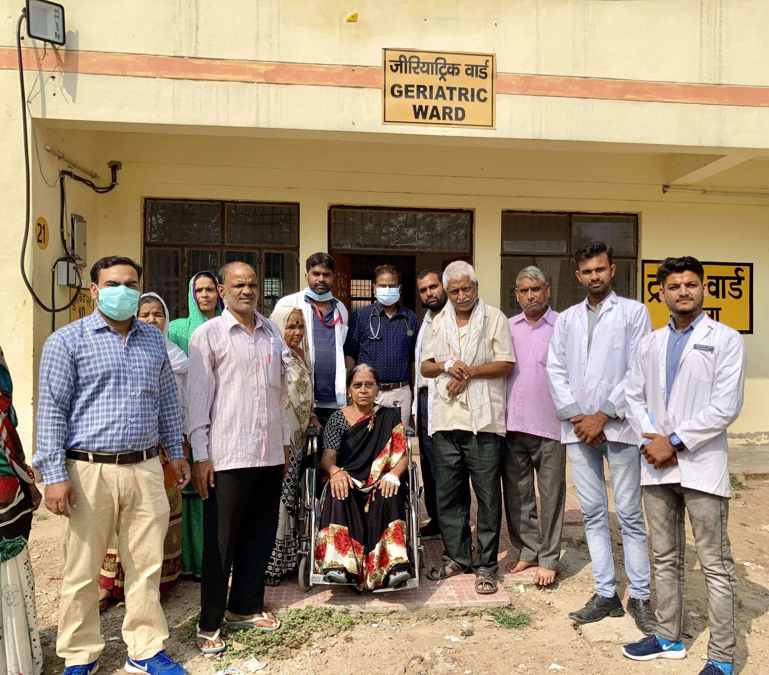
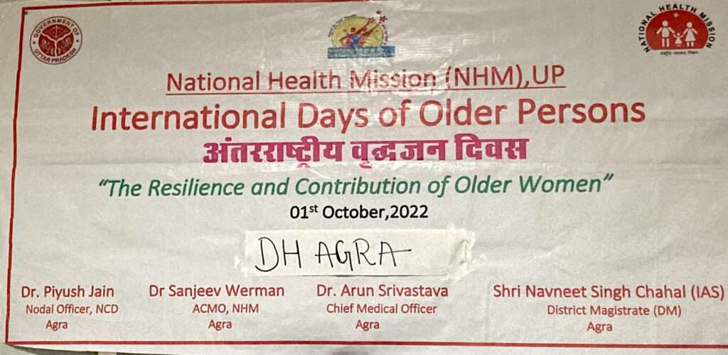
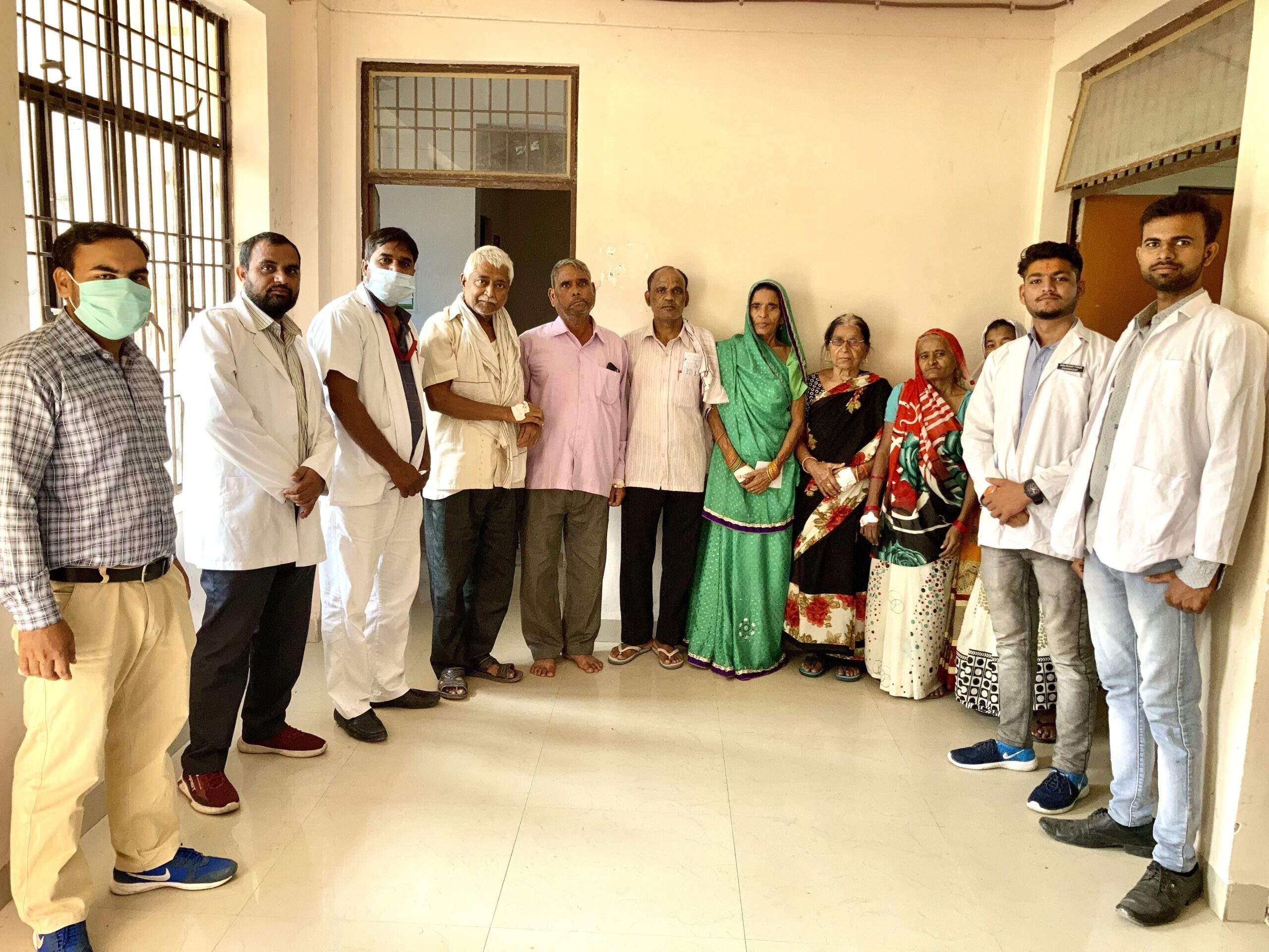
International Day of Older Persons 2022

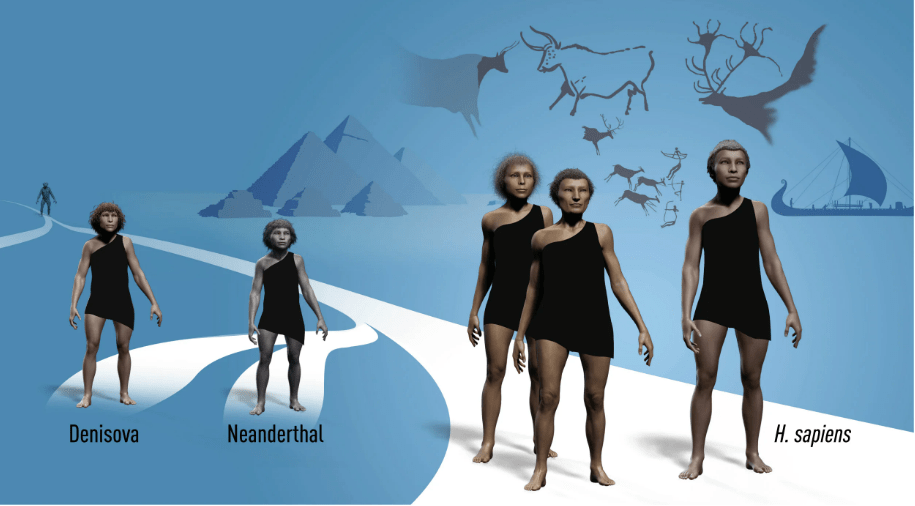
Theme of International Day of Older Persons: Resilience of Older Persons in a Changing World

The overall umbrella theme for the United Nations International Day of Older Persons in 2022 is “Resilience of Older Persons in a Changing World.” This theme will be celebrated by the NGO Committees on Ageing in New York, Geneva and Vienna – each with a unique and complementary approach to the overall theme.

Objectives

  • To highlight the resilience of older women in the face of environmental, social, economic and lifelong inequalities
  • To raise awareness of the importance of improved world-wide data collection, disaggregated by age and gender
  • To call on member states, UN entities, UN Women, and civil society to include older women in the center of all policies, ensuring gender equality as described in the Secretary-General’s report, Our Common Agenda

Common health conditions associated with ageing

Common conditions in older age include hearing loss, cataracts and refractive errors, back and neck pain and osteoarthritis, chronic obstructive pulmonary disease, diabetes, depression and dementia. As people age, they are more likely to experience several conditions at the same time.

Older age is also characterized by the emergence of several complex health states commonly called geriatric syndromes. They are often the consequence of multiple underlying factors and include frailty, urinary incontinence, falls, delirium and pressure ulcers.

International Day of Older Persons
International Day of Older person
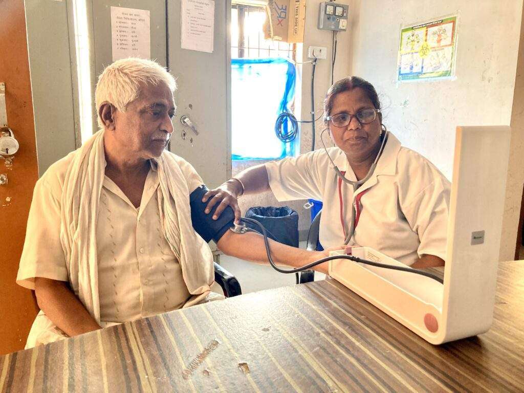
Blood Pressure
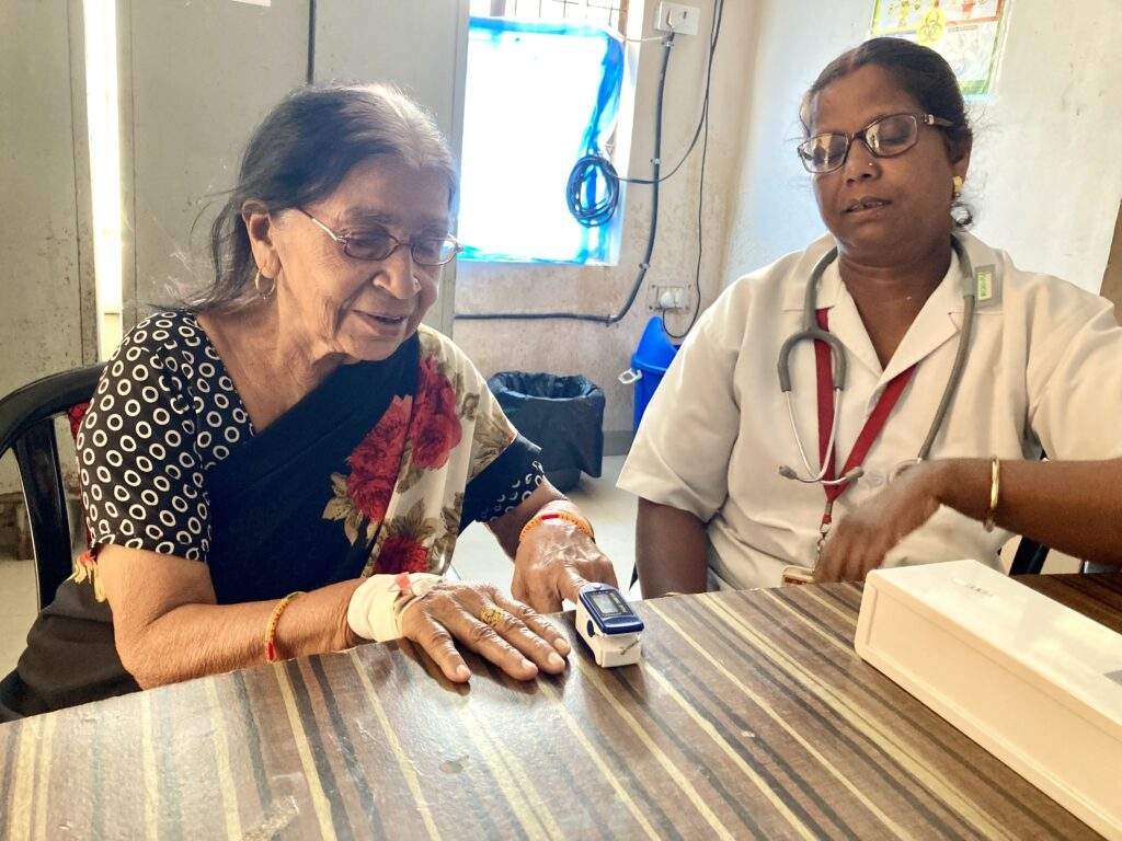
Pulse oximetry
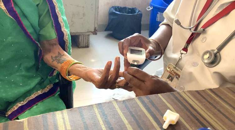
Glucometer
Dietary Advice
Nebulization
Comprehensive Geriatric Care

Background

On 14 December 1990, the United Nations General Assembly designated October 1 as the International Day of Older Persons (resolution 45/106). This was preceded by initiatives such as the Vienna International Plan of Action on Ageing, which was adopted by the 1982 World Assembly on Ageing and endorsed later that year by the UN General Assembly.

In 1991, the General Assembly adopted the United Nations Principles for Older Persons resolution 46/91. In 2002, the Second World Assembly on Ageing adopted the Madrid International Plan of Action on Ageing, to respond to the opportunities and challenges of population ageing in the 21st century and to promote the development of a society for all ages.

The composition of the world population has changed dramatically in recent decades. Between 1950 and 2010, life expectancy worldwide rose from 46 to 68 years. Globally, there were 703 million persons aged 65 or over in 2019. The region of Eastern and South-Eastern Asia was home to the largest number of older persons (261 million), followed by Europe and Northern America (over 200 million).

Over the next three decades, the number of older persons worldwide is projected to more than double, reaching more than 1.5 billion persons in 2050. All regions will see an increase in the size of the older population between 2019 and 2050. The largest increase (312 million) is projected to occur in Eastern and South-Eastern Asia, growing from 261 million in 2019 to 573 million in 2050.

The fastest increase in the number of older persons is expected in Northern Africa and Western Asia, rising from 29 million in 2019 to 96 million in 2050 (an increase of 226 per cent). The second fastest increase is projected for sub-Saharan Africa, where the population aged 65 or over could grow from 32 million in 2019 to 101 million in 2050 (218 per cent). By contrast, the increase is expected to be relatively small in Australia and New Zealand (84 per cent) and in Europe and Northern America (48%), regions where the population is already significantly older than in other parts of the world.

Among development groups, less developed countries excluding the least developed countries will be home to more than two-thirds of the world’s older population (1.1 billion) in 2050. Yet the fastest increase is projected to take place in the least developed countries, where the number of persons aged 65 or over could rise from 37 million in 2019 to 120 million in 2050 (225%).

Change the Way You Think About Age!